Tasks of the cerebrum
introduction
The cerebrum is the most widely known part of the brain. It is also called the endbrain or Telencephalon denotes and makes up most of the human brain. In this form and size it is only present in humans.
A rough distinction is made between four lobes on the cerebrum, which are named in relation to their anatomical position, and two separate, deep areas. More precisely, the cerebral cortex is called in 52 Brodmann areas divided, named after their first descriptor Korbinian Brodmann. It is divided into two halves called the hemispheres.
In order to have as large a surface as possible, it is folded many times. The windings and furrows that have arisen have their own names and can be assigned to specific functional areas.

General tasks of the cerebrum
The cerebrum is the highest instance of the central nervous system, which includes the brain as well as the spinal cord, and it is what makes people who they are with all their emotional, psychological and motor skills in the first place. It is involved in all active thoughts and movements, processes incoming information and then produces targeted answers and reactions. It is often linked to itself and other brain structures via nerve tracts. The nerve nuclei lie in the cerebral cortex and the nerve tracts in the medulla.
In addition to the anatomical classification, the cerebrum is functionally classified according to different aspects. This second division is based on the development and evolution of the brain. Parts of the human brain are also found in small mammals such as mice, while others are reserved for humans. One distinguishes the Paleocortex, the Striatum, the Archicortex and the Neocortex. They are all part of individual systems that are responsible for different areas of responsibility. However, they also work very closely together, which is why it is often not possible to draw clear boundaries between the individual areas.
The Paleocortex is the oldest part of the cerebrum. It is closely related to the olfactory brain and the sense of smell, the oldest of all the senses. It receives, transports and processes information that is captured by the olfactory organ, i.e. the sensory cells in the nose. That becomes him too Amygdala counted, an area that is responsible for emotional processes, especially for the development and processing of fear and anger. This also explains why smells can trigger such strong, emotional responses.
The Striatum sits deep inside the cerebrum and is part of the basal ganglia, a network of nerve cores and pathways that play an important role in the control of movement sequences.
It is also deeply seated Archicortex, which includes the hippocampus and is part of the limbic system. He is responsible for learning and memory processes. It has only recently become known that he is involved in spatial orientation. The limbic system as a whole is also responsible for life-sustaining functions such as the sex drive, food intake and the coordination of digestion.
The Neocortex is the youngest and by far the largest part of the cerebrum. The neocortex represents the actual surface of the cerebrum, which can also be viewed from the outside. In contrast to the previous structures, it does not lie in the depths of the brain. He is responsible for the collection of information from all areas of the body, as well as for the interpretation, association and transmission. It includes the motor centers for body movements, as well as the hearing, speech and vision centers. It is also the part of the brain that defines a person's personality. This part is called the as well Prefrontal cortex referred to because it lies far in front, directly behind the bony forehead. If this part of the neocortex is injured, massive personality changes and disorders result. Last but not least, it includes the areas of the brain that record sensory perceptions such as pain, vibrations and temperature differences.
Functions of the cerebral cortex
The cerebral cortex, also known as Cerebral cortex is visible from the outside and envelops the brain. It is also known as gray matter because when fixed it appears grayish in relation to the cerebral medulla. The cerebral cortex contains the nerve cores of the nerve tracts that move to other parts of the brain and the rest of the body.
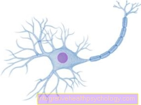
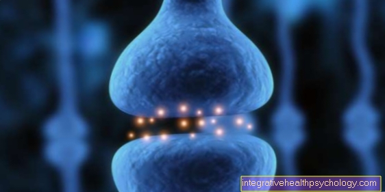
Here it is important to know the general structure of a nerve cell. Nerve cells or neurons consist of a cell body, an axon, which resembles a long process, and many dendrites. Dendrites are similar to antennas and receive signals from other nerve cells. This information is passed on to the cell body and processed there. This processed information can, in some cases, be forwarded for meters along the axon.
There are synapses at the end of the axon. They serve to pass on information to downstream nerve, muscle or gland cells. The cell bodies are collected and arranged in six layers in the cerebral cortex. They receive signals from the body in different layers than those from the rest of the brain. In this way a certain pre-sorting takes place. Depending on where the information comes from, it is forwarded to different other nerve cells.
The cerebral cortex serves, among other things, as a large transfer point for incoming stimuli and signals that have to be distributed to the right areas in order to ensure meaningful processing. It includes the two language centers. One is used to recognize and interpret spoken and written content. The second is responsible for the motoric and sensible production of language, i.e. words and sentences.
in the dorsal, that is, the rear part of the brain facing the back and the cerebral cortex is the center of vision. It is connected to other centers that interpret what is seen. Which of these centers the information is forwarded to depends, among other things, on the color of what is seen, whether it is moving or standing still. Faces are also interpreted in other places. The areas for facial recognition of other people and also of your own person are in turn connected to emotional centers, just to give an insight into the complexity of the cerebrum.
Of course, in the bark there is also a region for hearing. However, the largest part takes the so-called Motor cortex a. He is responsible for coordinating movements. To do this, he works closely with the somatosensory cortex together, which brings sensory impressions together. This also includes the Proprioception, also called depth perception. It provides information on the position of the muscles and joints in relation to one another so that the brain knows exactly where which muscle is located in order to then initiate and coordinate movements in a targeted manner. The senses also include the sense of touch, temperature, vibration and pain.
For the consciousness and the personality of a person is Prefrontal cortex responsible. It is closely linked to the memory and the emotional areas of the brain.
It is only the cerebral cortex that makes thinking in the form in which humans can operate possible and leads to us being aware of ourselves.

Functions of the cerebral medulla
The cerebral medulla is also known as white matter. It is made up of a network of supply and support cells, between which the nerve processes, the axons, run in bundles. These bundles are combined into lanes.
There are no cell bodies in the white matter. Your task is to sort the nerve tracts and supply them. Particularly large tracts are also known as fibers because they can be seen with the naked eye on the opened brain. They then, as the name suggests, look like fibers. Association fibers transport information within a cerebral hemisphere from one cortical area to the other, while commissure fibers connect the cortical areas of the two hemispheres with each other. Finally, a distinction is made between projection fibers that connect nerve nuclei in the cortex with nerve nuclei in the depths of the brain. These three fiber groups run exclusively in the cerebrum.
In addition, the cerebral cord contains pathways that lead into the cerebellum, the brain stem, the spinal cord and the extremities and in this way connect the cerebrum with other structures of the central and peripheral nervous system.
The cells in the cerebral medulla that are responsible for supplying and maintaining nerve cells are called glial cells. The glial cells of the central nervous system include the astrocytes, oligodendrocytes, microglia and ependymal cells.
The astrocytes mainly serve as supporting cells and are involved in building the blood-brain barrier. So they surround blood vessels that run in the brain and prevent pollutants and toxins from entering the brain.
Oligodendrocytes surround the long axons of nerve cells. In this way, they protect the axons, supply them with nutrients and isolate them. The insulation works in a similar way to ordinary electrical cables and ensures that information is passed on faster and more safely along the nerve processes.
As in the rest of the body, waste products from metabolic processes arise in the brain. These are absorbed and removed by the microglia.
Last but not least, there are the ependymal cells. They form a thin layer on the cerebral cortex, separating the cortex from the liquid spaces. The CSF spaces are filled with CSF, a liquid. The brain swims in this fluid. It is supplied and protected by the liquor and can give it off waste products, which are then transported into the body for disposal. Strictly speaking, the ependymal cells are not part of the cerebral cord, but are nevertheless counted among the supply cells of the central nervous system.
Tasks of the cerebral hemispheres and cerebral hemispheres
Although the two halves of the cerebrum look identical from the outside, they show certain differences in their function. They are divided into a dominant and a non-dominant hemisphere. By definition, the dominant hemisphere is that which processes motor and sensory language. While the sensory interpretation in the Wernicke Language Center takes place is that Broca area responsible for the formation and planning of words and sentences, i.e. the motor component of speaking. These two areas are therefore almost always in the dominant hemisphere. Interestingly, that is true Wernicke Center as the rational language center that leads to the understanding of a language.
In contrast, the language center for processing non-verbal, musical auditory impressions lies in the non-dominant half of the brain. For left-handers, the right hemisphere is usually the dominant one, for right-handers it is the left. This is because the motor and sensory functions of one half of the body are planned and interpreted in the opposite hemisphere.
In addition, the only one-sided on the non-dominant half comes posterior parietal cortex (= rear part of the lateral cerebral cortex). This is relevant for spatial orientation.

Cooperation of the cerebrum with the cerebellum
The cerebellum is located at the back of the skull, below the cerebrum. Also as Cerebellum known, it serves as a control center for the coordination, learning and fine-tuning of motion sequences. To do this, it receives information from the equilibrium organ in the ear, the spinal cord, the eyes, and the middle and cerebrum.
The cerebrum and the cerebellum work together when movement sequences are to be planned and carried out. Information always flows through intermediate structures and never directly from the cerebellum to the cerebellum or back. So the cerebrum is about so-called corticopontins Pathways connected to the pons, a structure in the brain stem. This then forwards the plans for a movement to the cerebellum. The cerebellum, in turn, works out the plans produced by the cerebral cortex and sends them back to the cerebral cortex via the thalamus.
The thalamus is located in the diencephalon and serves as a filter for incoming signals to the cerebrum.
The nerve pathways that run from the cerebrum to the cerebellum and vice versa cross each other on their way. This is relevant when determining disturbances in the movement sequence and must be taken into account in the diagnosis.


