Baker's cyst in a child
Introduction / definition
The Baker's cyst was first described in the 19th century by the English surgeon William M. Baker. It is also called Knee ganglion or Popliteal cyst designated. It is about a sack-shaped bulge of Bursa at the back of the knee joint, which is particularly common at Children up to the age of 15 years occurs. It often occurs without symptoms, but can cause a feeling of tension in the hollow of the knee.
In general, an observing and wait-and-see attitude applies in therapy, as the Baker's cyst can regress on its own in the child. Alternatively, surgery must be considered. The association with rheumatism is rare. An X-shaped misalignment of the legs is more common (Knock knees).
In addition to the real Baker's cyst in children, there is also the so-called clinical picture Pseudocyst. The cause is a joint disease or Bursitisas a result of which inflammatory fluid is formed. It can appear in the form of a bulging out of the bursa.
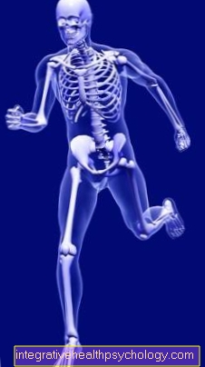
Anatomy knee joint

- Thigh muscles (Musculsus quadriceps femoris)
- Thighbones (Femur)
- Thigh tendon (Quadriceps tendon)
- Kneecap (patella)
- Kneecap tendon (Patellar tendon)
- Kneecap tendon attachment (Tibial tuberosity)
- Shin (Tibia)
- Fibula (Fibula)

I would be happy to advise you!
Who am I?
My name is I am a specialist in orthopedics and the founder of .
Various television programs and print media report regularly about my work. On HR television you can see me every 6 weeks live on "Hallo Hessen".
But now enough is indicated ;-)
The knee joint is one of the joints with the greatest stress.
Therefore, the treatment of the knee joint (e.g. meniscus tear, cartilage damage, cruciate ligament damage, runner's knee, etc.) requires a lot of experience.
I treat a wide variety of knee diseases in a conservative way.
The aim of any treatment is treatment without surgery.
Which therapy achieves the best results in the long term can only be determined after looking at all of the information (Examination, X-ray, ultrasound, MRI, etc.) be assessed.
You can find me in:
- - your orthopedic surgeon
14
Directly to the online appointment arrangement
Unfortunately, it is currently only possible to make an appointment with private health insurers. I hope for your understanding!
Further information about myself can be found at
Symptoms
In most cases, the Baker's cyst hardly causes any discomfort in the child. Pain can be radiated into the thigh region, the back of the knee and the muscles of the calves. More common, on the other hand, is a feeling of tension in the hollow of the knee, which increases with physical exertion and then decreases again. This is due to an increased volume of fluid in the cyst. Usually the size of the cyst varies between a walnut and a chicken egg.
Movement restrictions can occur particularly when bending the knee joint. The larger the cyst, the more severe the symptoms in the form of pain and restricted mobility. When the cyst is felt, a firm, elastic structure dominates, which should not be confused with a tumor.
A possible complication is the bursting of the cyst, in which the fluid contained in it leaks into the tissue. The symptoms show up with increasing swelling and pressure-dependent pain. The symptoms are similar to the symptoms of deep vein thrombosis, which should be diagnosed. If the fluid-filled tissue sac exerts pressure on a nerve, sensory disturbances up to paralysis can occur.
root cause
The exact cause of the Baker's cyst in children has not yet been clarified. The sack-shaped bulge occurs spontaneously. One assumes an innate Overproduction of synovial fluid from who are the Path of Least Resistance seeks. Since the joint capsule on the back of the knee joint is particularly flexible, a spherical bulge develops at this point. It pushes through the head of the muscle Calf muscle (M. gastrocnemicus) and the tendon attachment of the semimembranous muscle (M. semimembranosus). They occur more frequently in boys under the age of 15.
In contrast to the adult pseudocyst, the Baker's cyst is less associated with diseases of the rheumatic type than with an X-shaped deformity of the legs. Underlying diseases such as rheumatism and arthrosis are therefore not among the causes in childhood.
In children, a so-called ganglion can develop as a result of permanent irritation of the muscular tendon sheaths.
Diagnosis of a Baker's cyst in a child
The diagnosis can be based on the tactile findings, the symptoms and a Ultrasound examination put. This relatively simple procedure is usually sufficient for children. From a diameter of two centimeters the tactile finding is clear. Smaller variants can also be used in ultrasound, but also in Magnetic resonance imaging (MRI) can be detected. The ultrasound provides information about the volume and spread of the cyst. MRI is rarely used to diagnose classic Baker's cysts.
In the case of the pseudocyst, it provides additional information on the underlying disease and signs of wear and tear. However, an MRI scan can help differentiate a Baker's cyst from a sarcoma be helpful.If there is a radiological suspicion of a sarcoma, the diagnosis is confirmed with a tissue sample. A malignant occurrence in the form of a tumor, hematoma, bulging of the veins and thromboses should be excluded in any case by differential diagnosis.
Therapy of a Baker's cyst in a child
In many cases, the Baker's cyst regresses on its own in the child and does not require any further therapy. Conservative measures include taking it anti-inflammatory drugs. Cortisone Preparations are controversial in their application. Particularly large cysts can be used Puncture emptied if there are existing symptoms. These can be restricted mobility, symptoms of paralysis or pain. To do this, the attending physician pierces the sac under sterile conditions and removes the liquid it contains.
Alternatively there is the possibility of operative removal. The operation of a Baker's cyst in children is only considered in rare cases. After the cyst has been surgically exposed, the connection between the cyst and the joint capsule is ligated. Then it is cut out and the Sewn capsule. This serves as a preventative measure for cyst formation again. After the operation, the leg is raised and cooled. In addition, a plaster of paris or a splint is used for immobilization. Passive mobilization begins after three days and active movement of the knee joint takes place after just seven days. The minimally invasive technique is not used in children. The cyst reappears after every tenth operation. While the causes in adults have been clarified, there is still no adequate explanation for the recurrence of Baker's cyst in children.
forecast
Generally, the prognosis of Baker's cyst in children is Well. It often disappears spontaneously in childhood and one can choose an observational approach. As a therapeutic measure for large cysts, puncture does not guarantee that the cyst will disappear. A recurrence should be expected.
prophylaxis
Since the Baker's cyst is congenital in children, it cannot be prevented.


