The pulmonary circulation
General
As a pulmonary circulation (small cycle) is the term used to describe the transport of blood between the lungs and the heart. It is used to enrich the oxygen-poor blood from the right heart with oxygen and to transport the oxygen-rich blood back to the left heart.
From there, the oxygen-rich blood is pumped back into the body. Although the pulmonary vessels contain a lot of oxygen, the lungs still need their own vessels to supply themselves with oxygen. In order to differentiate between the two vascular circuits, the lungs' own vessels are called Vasa privata designated. The oxygenation vessels for the rest of the body are those Vasa publica.

The function of the pulmonary circulation
The function of the pulmonary circulation is to transport the blood between the heart and the lungs. It is used for gas exchange, which means the renewed uptake of oxygen into the blood and carbon dioxide release via the breath. The gas exchange takes place in the Alveoli (Alveoli) instead.
During breathing, carbon dioxide (CO2) is released via diffusion and oxygen (O2) is absorbed from the alveolar air into the blood. So that the oxygen can be transported in the blood, it is bound to the red blood pigment hemoglobin. The blood is now rich in oxygen (= oxygenated) and is transported back to the heart via a venous system. The oxygen-rich blood is then transported from there to the rest of the organs in the body via what is known as the large body circulation.
The vessels of the pulmonary circulation are called Vasa publica, as it enables gas exchange and this serves the entire organism. In contrast to this, the vessels that supply the lungs themselves are called Vasa privata.
You might also be interested in this topic: The human bloodstream
Vasa publica
The oxygen-poor blood from the body first arrives via the two large vena cava (Superior vena cava and inferior) in the right atrium of the heart.
During diastole, the tricuspid valve, which separates the right atrium and the right ventricle, opens, and the oxygen-depleted blood enters the right ventricle.
In the expulsion phase of the heart (systole), the blood is drawn through the large pulmonary trunk (Pulmonary trunk) discharged into the vessels of the lungs. This large trunk divides into the right and left large pulmonary arteries (Pulmonary artery). This artery branches into the smallest capillaries in the respective lungs. In this capillary area of the alveoli (Alveoli) the gas exchange takes place.
The CO2 produced in the body is released from the blood and exhaled, while the oxygen-containing air is absorbed into the smallest bronchi when inhaled and can get into the blood via the alveoli.
The oxygen-containing blood now flows back to the heart in various pulmonary veins. In this way, the smallest veins join together to form increasingly larger veins, until finally the left and right large pulmonary veins (Pulmonary vein) open into the left atrium. From there, the oxygen-rich blood reaches the left ventricle (left ventricle) via the mitral valve during diastole. During the expulsion phase of the heart (systole), the now oxygen-rich blood is pumped through the aortic valve into the aorta and thus the large body circulation.
Vasa privata
Since the walls of the bronchi are too thick and the air flow rate too high, the lungs need their own vessels to supply them.
The small branches of these vessels are called the rami bronchiales.
The bronchial rami of the left lung arise from the thoracic artery, the vessels of the right lung also originate from the various vessels in the intercostal spaces (Intercostal artery).
The venous drainage of these arteries reaches the azygos vein on the right side near the hilus and the hemiazygos vein on the left. The peripheral small veins (venae bronchiales) open into the large pulmonary veins of the vasa publica.
- Pulmonary blood flow
- Vascular supply to the lungs
anatomy
The pulmonary circulation starts in the right part of the heart. The blood that supplied the organs with oxygen is now enriched with carbon dioxide and low in oxygen. This blood from the body is drawn through the right atrium and the right main chamber (= Ventricle) in the Pulmonary trunk (= Pulmonary artery) pumped.
The pulmonary trunk divides into a right and a left pulmonary artery along the anatomy of the airways. These branch out into ever smaller vessels up to the so-called capillaries. They surround the many millions of alveoli (Alveoli) that are filled with air. The blood flows very slowly in the capillaries because this is where the oxygen exchange takes place between the alveoli and the capillaries. The carbon dioxide is released through the thin walls of the capillaries and alveoli and exhaled through the breath, while oxygen is absorbed into the bloodstream in return.
The smallest veins, so-called venules, unite from the capillaries to form increasingly larger veins and transport the now oxygen-rich (= oxygenated) Blood back to the heart. It now reaches the left part of the heart and is pumped from there via the aorta into the body's circulation.
Also read our topics:
- The vessels of man
- The cardiovascular system
Change of circulation at birth
This pulmonary circulation is not required before birth, as the fetus is supplied with oxygen from the mother via the umbilical cord. The lungs are not yet ventilated. For this reason there is between the Pulmonary trunk and the aorta has an opening called the ductus arteriosus. There is also a small hole between the right and left atrium (Foramen oval).
With the first cry after the birth, the pressure conditions are reversed because the lungs are ventilated. Now both that Foramen oval, as well as the Ductus arteriosus shut down. If this does not happen, various problems of adaptation can arise in the newborn and therapy or surgery to close the opening may be necessary.
What is the pressure in the pulmonary circulation?
The pulmonary circulation is part of the so-called low-pressure system. The average pressure is between 0 and 15 mmHg. The low pressure system includes the Capillaries, the Veins, the right part of the heart, the vessels of the Pulmonary circulation and the left atrium of the heart.
In the body's circulation, on the other hand, as part of the so-called high-pressure system, pressures between 70 and 120 mmHg prevail at rest.
All vessels of the low pressure system are characterized by a higher elasticity than the vessels of the high pressure system. The reason for this lies in the main task of the low-pressure system - the intermediate storage of the blood. If there is a shortage of blood and, as a result, an insufficient supply of blood to the organs, the blood volume stored in the low-pressure system can be used to initially ensure a supply of the organs.
Pulmonary circulatory disorders
The pulmonary embolism
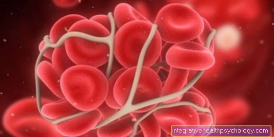
A pulmonary embolism is a narrowing or complete blockage (occlusion) of a pulmonary artery or bronchial artery by one Embolus.
An embolus is an endogenous or foreign object that causes a narrowing of the vascular system (= embolism) leads. There are different forms of pulmonary embolism, the main cause being thromboembolism.
About 90% of the embolus is a detached thrombus, e.g. a clot from a deep leg vein, but it can also come from other vessels. A pulmonary embolism can be life-threatening under certain circumstances, as it results in a restricted supply of oxygen.
Furthermore, the right heart is subjected to excessive stress, as it has to pump against increased pressure due to the narrowing of the vessels. A so-called cor pulmonale develops. The heart's pumping capacity is insufficient. This means that the lungs are no longer adequately supplied with blood and, as a result, the organism no longer receives sufficient oxygen.
Pulmonary embolism can manifest itself as chest pain, increased breathing rate, and shortness of breath. Furthermore, the heart rate is greatly increased and symptoms such as dizziness, sweating and fever can occur. Not all symptoms can be determined with certainty in a pulmonary embolism. In addition to imaging procedures (X-ray, CT), an EKG and / or an echocardiography are usually also performed.
Pulmonary embolism therapy depends on the severity of the embolism. In most cases, anticoagulants are used (= Blood thinner) administered to prevent the formation of new thrombi. The existing thrombus is usually removed by means of lysis therapy, which means using drugs that dissolve the thrombus. In severe cases, the thrombus can also be removed using a right heart catheter or surgery.
Learn more about this at: Causes of pulmonary embolism
Anatomy of air ducts

- Right lung -
Pulmodexter - Left lung -
Pulmo sinister - Nasal cavity - Cavitas nasi
- Oral cavity - Cavitas oris
- Throat - Pharynx
- Larynx - larynx
- Trachea (approx. 20 cm) - Trachea
- Bifurcation of the trachea -
Bifurcatio tracheae - Right main bronchus -
Bronchus principalis dexter - Left main bronchus -
Bronchus principalis sinister - Tip of the lung - Apex pulmonis
- Upper lobe - Superior lobe
- Inclined lung cleft -
Fissura obliqua - Lower lobe -
Inferior lobe - Lower edge of the lung -
Margo inferior - Middle lobe -
Lobe medius
(only on the right lung) - Horizontal cleft lung
(between the upper and middle lobes on the right) -
Horizontal fissure
You can find an overview of all Dr-Gumpert images at: medical illustrations

- Bronchiole
(cartilage-free smaller
Bronchus) -
Bronchiolus - Branch of the pulmonary artery -
Pulmonary artery - End bronchiole -
Respiratory bronchiolus - Alveolar duct -
Alveolar duct - Alveoli sheath -
Interalveolar septum - Elastic fiber basket
of the alveoli -
Fibrae elasticae - Pulmonary capillary network -
Rete capillare - Branch of a pulmonary vein -
Pulmonary vein
You can find an overview of all Dr-Gumpert images at: medical illustrations
Summary
The pulmonary circulation describes the small circulation between the lungs and the heart, which enriches the oxygen-poor blood and transports the oxygen-rich blood back to the heart. Since the lungs themselves also need an oxygen supply, the vessels of the lungs are divided into Vasa privata and Vasa publica. This cycle is not needed before the birth, only with the first cry does the pressure change and the small cycle is set in motion.
Recommendations from our editorial team
- Pulmonary blood flow
- Vascular supply to the lungs
- Cardiovascular system
- water in the lungs
- Fallot tetralogy


