The thoracic spine
Synonyms
Thoracic vertebrae, thoracic vertebrae, kyphosis, dorsalgia, rib blockage, vertebral blockage
anatomy
The thoracic spine is part of the spine as a whole, also known as the backbone. There are 12 thoracic vertebrae (vertebrae thoracicae), which make up the middle part of the spine and together with the ribs (costae) and the breastbone (sternum) form the chest (thorax).
The thoracic spine naturally has a slight curvature when viewed from the side (kyphosis). The spine is curved convexly back here. If the thoracic spine curvature is pathologically increased, for example in Scheuermann's disease or osteoporosis, there is a hunched back (hyperkyphosis), also known as a “hump” as an extreme form. Lateral deviation of the spine is known as scoliosis.

Figure thoracic spine

Thoracic spine (green)
- First cervical vertebra (carrier) -
Atlas - Second cervical vertebra (turner) -
Axis - Seventh cervical vertebra -
Vertebra prominent - First thoracic vertebra -
Vertebra thoracica I - Twelfth thoracic vertebra -
Vertebra thoracica XII - First lumbar vertebra -
Vertebra lumbalis I - Fifth lumbar vertebra -
Vertebra lumbalis V - Lumbar cruciate ligament kink -
Promontory - Sacrum - Sacrum
- Tailbone - Os coccygis
You can find an overview of all Dr-Gumpert images at: medical illustrations
Obstructions in the thoracic spine
In the area of the thoracic spine, blockages of the vertebral joints or the costal vertebral joints often arise. Typically, a pain is felt between the shoulder blades, which can also pull in a belt-shaped manner towards the chest. The pain associated with a blockage is mostly movement-dependent, sometimes breathing-dependent.
The vertebral body sits back in the Vertebral arch away. The vertebral arches beginning in the vertebral body are called arch roots (pedicles). They are of particular importance in spinal surgery because, within the framework of the instrumentation, they enable the screws to stiffen the spine (Spinal fusion) in the Vertebral bodies be introduced. The posterior end of the vertebral arch forms the vertebral plate (lamina). The begins where the two vortex plates meet Spinous process (Processus spinosus), which protrudes steeply downwards in the area of the thoracic spine and is easy to feel on the back, even for the layman. The vertebral arch, together with the vertebral body, forms the vertebral hole (foramen vertebrale, vertebral canal, spinal canal) through which the spinal cord pulls towards the foot. The clinical picture of Spinal stenosis describes a spinal canal that is too narrow with resulting nerve damage to the spinal cord or the Spinal nerves.
In the side view of the Spine The arch roots of two adjacent vertebral bodies each form a hole open to the side (foramen intervetebrale, neuroforamen), from which the spinal cord nerves (spinal nerves) leave the vertebral canal.
In addition to the spinous process, the transverse processes (processus transversi) also extend from the vertebral arch, which serve as starting points for the muscles accompanying the spine and the ligamentous structures.
Two further smaller processes above and below form the upper and lower intervertebral joint (superior and inferior articular processes; facets). They are important for the mobility of the thoracic spine. With wear and tear of the articulated cartilage surface, stubborn back pain can arise, known as symptomatic spondylarthrosis or as Facet syndrome are designated. However, the most common occurrences of facet syndrome are Lumbar spine and the cervical spine.
As a specialty of the Thoracic spine the articulated connection to the ribs can be seen. Together with the Ribs and the sternum (sternum), a kind of cone-shaped basket that is open at the top and bottom (upper and lower thoracic aperture) is formed, which is why the Rib cage (thorax) speaks.
The joint socket for the head of the ribs is located on the upper and lower edges of the thoracic vertebrae.
The individual thoracic vertebra consists of a thoracic vertebral body (corpus vertebra), a thoracic vertebral arch (arcus vertebra) and thoracic vertebral processes (processus vertebrae).
The Vertebral bodies consists mainly of spongy bones (cancellous bone). Its fixed bone ends (cortex) facing downwards and upwards are also known as base and cover plates. The neighboring one lies on top of them Intervertebral disc, then the next vertebral body follows. The thickened lateral edges of the vertebral bodies are called the marginal ridges.
These terms have a practical meaning especially when describing changes in the roentgen or MRI of the thoracic spine.

CT image of the spine
- Vertebral bodies
- Transverse process
- Articular process / vertebral joint
- Spinous process
- Vertebral hole
Function of the thoracic spine
The range of motion of the thoracic spine is small, as the attachment of the ribs and the roof-tile-like arrangement of the spinous processes do not allow a great deal of freedom of movement. The most important function of the thoracic spine is to rotate the trunk. Rotational movements of the trunk are almost invariably carried out in the lower thoracic spine.
In addition, the movements of the rib-vertebral joints are important for inhalation and exhalation. Due to a different type of joint, the upper pairs of ribs of the 2nd-5th Rib (swivel joint) a different rib movement (forward lifting of the ribs) than the pairs of ribs of the 6th-9th centuries. Rib (sliding joint; lateral lifting of the ribs). Overall, this increases the volume of the chest when inhaling. Diseases that limit this mobility (e.g. ankylosing spondylitis) lead to the disruption of breathing movements (breathing excursion).
The smallest functional (movable) unit of the spine is that Motion segment. A movement segment is understood to be the unit between two adjacent vertebral bodies, which are connected to one another via two vertebral joints, as well as the one between the vertebral bodies Intervertebral disc and all muscular structures, ligament and nerve structures that are in this area.

Figure spine sections
The red colored area shows the different sections of the spine.
Left to right:
- Cervical spine and upper thoracic spine
- Thoracic spine
- Lumbar spine

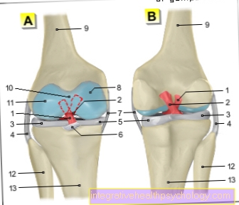
Side view motion segment
- Vertebral bodies
- Intervertebral disc
- Spinal nerve root
- Intervertebral hole (neuro foramen)
- Vertebral joint
- Spinous process of the vertebra (palpable on the back as the rear end of the vertebra)
Isolated disorders are often found in a single movement segment (e.g. Blockages, Herniated disc of the thoracic spine). For a local description of a spinal disease, the individual vertebral bodies are counted, e.g. HWK 5 for the 5th cervical vertebrae, BWK 9 for the 9th. Thoracic vertebrae, LWK 3 for the 3rd lumbar vertebral body, etc. The same applies to the intervertebral discs and the movement segments. The description BWK 7/8 refers to the movement segment between the 7th and 8th thoracic vertebrae.
In addition to its function as a static organ and as an organ of movement, the spine also has another important function as a protective and conducting organ for the spinal cord. The spinal cord basically represents the extension of the brain and is therefore also the central nervous system assigned.

Figure spine
- Transverse process
- outgoing nerve
- Vertebral bodies
- Spinous process
- Spinal cord
Appointment with a back specialist?

I would be happy to advise you!
Who am I?
My name is I am a specialist in orthopedics and the founder of .
Various television programs and print media report regularly about my work. On HR television you can see me live every 6 weeks on "Hallo Hessen".
But now enough is indicated ;-)
The spine is difficult to treat. On the one hand it is exposed to high mechanical loads, on the other hand it has great mobility.
The treatment of the spine (e.g. herniated disc, facet syndrome, foramen stenosis, etc.) therefore requires a lot of experience.
I focus on a wide variety of diseases of the spine.
The aim of all treatment is treatment without surgery.
Which therapy achieves the best results in the long term can only be determined after looking at all of the information (Examination, X-ray, ultrasound, MRI, etc.) be assessed.
You can find me in:
- - your orthopedic surgeon
14
Directly to the online appointment arrangement
Unfortunately, it is currently only possible to make an appointment with private health insurers. I hope for your understanding!
You can find more information about me at
Thoracic spine disorders
Diseases of the thoracic spine are less common than in the area of the cervical and lumbar spine, especially with regard to the frequent wear-related diseases (e.g. Facet syndrome, herniated disc, spinal canal stenosis Etc.).
Still, dorsalgias come as one Back pain unspecifically designated in the thoracic spine, often before. In young people, this often conceals blockages of the intervertebral joints or the rib-vertebral joints. A blockage is understood as the temporary, reversible reduced mobility of a joint, which causes pain due to the "hooked" deformity and the capsular tension of the joint. Muscle tension of any kind can arise independently, for example due to incorrect loading, or appear as a concomitant disease.
A Herniated disc of the thoracic spine is very rare (<1% of all Herniated discs).
In the elderly, thoracic spine pain is often caused by a decrease in bone mass (osteoporosis) caused. This disease, which is already painful in itself, can become more pronounced in the thoracic spine if it becomes one Vertebral fracture due to the severely reduced carrying capacity of the vertebral body has come. Modern minimally invasive therapy methods such as the Vertebroplasty and the Kyphoplasty, are used more and more often to treat such breaks (fractures).
Accident-related (traumatic) vertebral body fractures occur more frequently in the area of the transition from the thoracic to the lumbar spine. The main reason for this is the reversal of curvature from thoracic spinal kyphosis to lumbar lordosis.
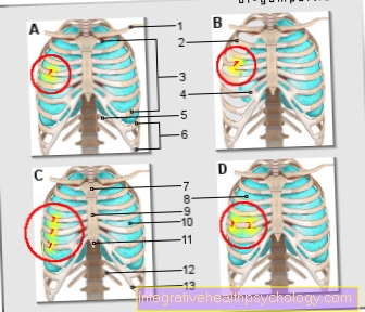
Thoracic spine kinesiotape
Taping describes colloquially the creation of a Tape association. The material used here is wide adhesive strips that are available in numerous colors today.
The aim of a tape association is that targeted restriction of mobility of the desired Joint while maintaining a Residual function and thus one Residual mobility.
This can happen with some Diseases or Injuries the Healing process accelerate compared to a complete immobilization. Tape dressings can therefore only be used in areas where complete immobilization (Immobilization) necessary is.
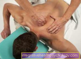
A large area of application for tape bandages are Pulled muscles.
A tape dressing can do both therapeutic - so after an injury - as well prophylactic - that is, as a preventive measure.
For pain in the area of the Thoracic spine Usually only so-called kinesio tapes are used. A kinesio tape of the thoracic spine can be one Relieving pain procure. Before doing this, however, the root cause for the Pain through a doctor to be clarified. Depending on the location of the Maximum pain and depending on Art there are different ways to tape the bandage, the assistance and guidance of a Physiotherapists is an advantage here.
Read more on this topic at: Kinesio tape
Herniated disc of the thoracic spine

Where one vertebral body meets the next, the intervertebral disc (Discus intervetebralis) acts as a kind of buffer zone. It lies between the two vertebral bodies and consists of a fibrous outer ring (annulus fibrosus) and an inner part filled with gelatinous mass (nucleus pulposus).
The inner mass has a high water-binding capacity and thus serves as a kind of water cushion for the spine.
Over the course of life, the limiting outer ring can develop more and more small cracks. In the worst case, this leads to a tearing and an exit of the nucleus pulposus towards the outside in the direction of the spinal cord. This is known as a herniated disc, disc prolapse, disc prolapse or disc herniation.
One of the most common causes for this is years of incorrect strain, even people who mainly sit in everyday life seem to be more often affected by a herniated disc. Of the 23 intervertebral discs that the human body has, those in the lumbar spine area are by far the most frequently affected by a herniated disc. Such an event rarely occurs in the thoracic spine area. Depending on the source, 0.2-5% of all herniated discs are said to be located in the thoracic spine (thoracic spine).
It is not uncommon for such a thoracic disc prolapse to be an incidental finding in magnetic resonance imaging (MRI of the thoracic spine) because initially it does not cause any symptoms. But it can also be noticed by unspecific pain in the thoracic spine area. These can radiate down the course of the ribs and also towards the heart and abdominal wall and can thus be misinterpreted under certain circumstances.
It is not uncommon for an inflammation of the gallbladder, ulcer in the stomach or small intestine, an inflammation of the esophagus or an inflammation of the kidney to be considered first.
Sensitive failures in the sense of numbness or abnormal sensations as well as motor function restrictions in the sense of a weakened muscle function of the affected muscles can occur, but are less common first symptoms than pain.
Symptoms of a thoracic disc herniation can also be disorders of the bladder function or the anal sphincter. The thoracic disc herniation mostly affects the lower part of the thoracic spine and has a maximum frequency between the ages of 40 and 50.
It can slowly increase over the years, so it can have a chronic course but can also begin very acutely.
If a herniated disc of the thoracic spine is suspected, imaging of the spine using an MRI of the thoracic spine (magnetic resonance imaging) is the method of choice. A prolapse can usually be easily recognized here. If the diagnosis is confirmed, a therapeutic approach must be decided. In the case of herniated discs of the thoracic spine, conservative - i.e. non-operative - therapy is often sufficient.
Heat applications and pain relievers are used here. It is important - if the pain allows it - to move sufficiently, physical protection can possibly cause further damage here. However, this depends on the individual case.
In the long run, attending a back school is a helpful method to learn how to deal with your own spine correctly and to avoid the symptoms flaring up again.
Sports that are gentle on the intervertebral discs, such as swimming, hiking and Nordic walking, promote the recovery process and can prevent further complaints. Thoracic prolapse rarely has to be treated surgically. This is the case when sensory or motor deficits or disorders of bladder or rectal function occur or have occurred. Here - depending on the severity - rapid action may be required.
Read more on this topic at:
- Herniated disc of the thoracic spine
and - MRI for a herniated disc
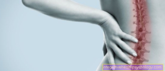
Thoracic spine pain
Since the thoracic spine is relatively immobile compared to the cervical and lumbar spine, pain is rare here. Nevertheless, pain in other locations can radiate here and thus simulate a disorder in the thoracic spine area.
In the field of manual medicine (chiropractic), pain in the spine is often referred to as "blockages". This should result in a temporarily restricted mobility within the spine, which, however, exists without any specific organic change. So one cannot find a presentable cause for such a functional restriction. Therefore, the concept of blocking is not without controversy among medical professionals.
Blockages often lead to subsequent muscle tension, which in turn leads to pain.
In manual therapy, deblocking by means of special handles is an option. In conventional medicine, treatment with physiotherapy, warmth and painkillers is usually indicated for thoracic spine pain of this type.
Another possible cause of pain in the thoracic spine are degenerative changes, so to speak, age-typical signs of wear and tear, which are more pronounced in some people and less in others. However, they are very rare in the thoracic spine area.
Intercostal neuralgia, also called intercostal neuralgia, can cause belt-shaped pain in the thoracic spine that radiates along the ribs.
The pain is caused by irritation of the nerves that run below each rib. The pain is often accompanied by a sensory disturbance in the area of the affected nerves. Therapy is mostly with painkillers.
Other typical diseases that directly affect the spine and can lead to pain include scoliosis, a congenital malformation of the spine or ankylosing spondylitis (ankylosing spondylitis), a rheumatic disease that occurs more frequently at a young age. Also typical is the slipped disc of the thoracic spine, which manifests itself between the ages of 40 and 50. The latter two diseases, however, more often affect the cervical or lumbar spine or the sacral part of the vertebral system.
There are other non-orthopedic but very important differential diagnoses that should be considered in the case of an acute onset of pain in the thoracic spine:
In the case of a pneumothorax, air incorrectly enters the pleural space due to a defect in the pleura (pleura).
In the most spectacular case, this can be done by stabbing a knife, for example.
But such a defect can also originate inside the body, this is more common in young, tall men. If air gets into the pleural space, there is no longer any negative pressure.
As a result, the affected side's lungs contract and are barely available for breathing.
This can quickly develop into a life-threatening condition, especially if it is a special form of pneumothorax, the tension pneumothorax.
Here, air can no longer get into the pleural space, so that one side of the chest continues to inflate and important structures such as the windpipe and the heart are displaced.
In addition to chest pain, shortness of breath, accelerated breathing (tachypnea) and irritation of the throat can occur. Tension pneumothorax can also cause an accelerated heartbeat (tachycardia) and falling blood pressure.
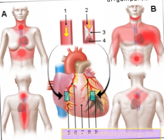
An X-ray is the diagnostic tool of choice. If there is pain in the thoracic spine, a heart attack should also be ruled out. In the case of a heart attack, the pain typically radiates from the left side of the chest into the left arm, but there are numerous other possible pain locations such as the right arm, lower jaw, upper abdomen and back.
An EKG and a determination of the cardiac enzymes by means of a blood sample are important diagnostic tools here.
Disturbances in the area of the gallbladder such as gallstones (chole (cysto) lithiasis) or inflammation of the gallbladder (cholecystitis) can cause pain in the shoulder and upper back area. In rare cases, pain caused by inflammation of the pancreas (pancreatitis) can also reach the upper back.
Read more on this topic at: Thoracic spine pain
Fracture of the thoracic spine

A fracture (fracture) of the thoracic spine can have various causes. Such injuries are common in elderly patients, especially women with osteoporosis. The bones here become significantly more brittle and fragile due to degradation processes, so that a fracture in osteoporosis often occurs without actual trauma.
Here the pain symptoms are sometimes significantly less acute than in fractures that result from violence. Sometimes such so-called osteoporotic sintering fractures are incidental findings during an X-ray of the spine.
A classic example of a vertebral fracture caused by trauma is diving into the water that is too shallow. Here, however, the cervical spine is more often affected than the thoracic spine.
Other accidents such as falls while exercising or traffic accidents can cause spinal fractures.
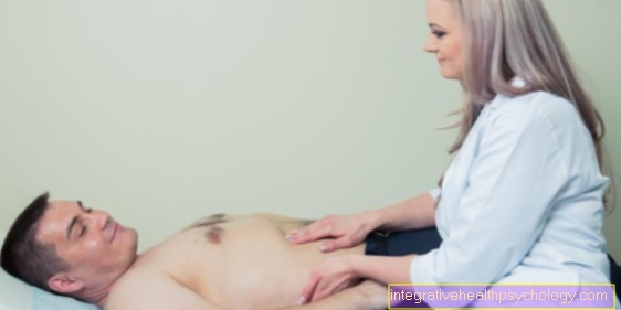
In the case of fractures caused by such trauma, there is usually significant pain; on examination, pressure or tapping pain in the affected section of the spine is typical.
Regardless of the cause, the method of choice for imaging is always an X-ray examination of the spine, usually in two planes, i.e. from the front and from the side. A precise neurological examination is always important in the event of an injury to the spine, since - depending on which segment or which segments of the vertebra is / are affected by the fracture - a lesion of the spinal cord can occur which runs behind the vertebrae. Involvement of the spinal cord can be indicated by sensory or motor deficits or disorders of the bladder or rectal function.
In this case, it is essential to act quickly, as otherwise, in the worst case, there may be cross-sectional symptoms.
But even if the spinal cord is not affected, untreated healed vertebral body fractures can lead to symptoms such as chronic pain or misalignments. Treatment for a thoracic spine fracture depends, among other things, on the type of fracture and the age of the patient and the degree of impairment. Fractures in this area can be treated both conservatively and surgically.
In the case of conservative measures, the focus is primarily on pain therapy and physiotherapeutic treatment; depending on the fracture, a corset can be used to stabilize the fracture from the outside. If surgical treatment of a fracture is necessary, a so-called internal fixator is often used, a kind of metal framework that connects several vertebrae with inserted screws and rods and thus stabilizes the broken vertebra; this is also known as spondylodesis.
This procedure results in a stiffening of the affected vertebral segments, i.e. a restriction of mobility. However, it is a good procedure, especially in the thoracic spine area, since the range of motion that is possible here is by nature not too great.
The so-called kyphoplasty is another surgical procedure. It can be used for stable fractures, i.e. those in which the spinal cord is not endangered; the vertebral body is straightened up again by introducing material. This procedure is more commonly used for osteoporotic vertebral fractures.
Radiation to the stomach
Lesions in the area of Thoracic spine can cause discomfort that extends into the Radiate epigastric region.
But also stomach problems, for example Ulcers, can cause discomfort that extends in the area of the Thoracic spine noticeable and so falsely lead to the assumption that the cause of the discomfort is in the area of Spine must be sought.
Therefore, in the event of complaints in the area of Spine It should be borne in mind that the cause of this is also in internal organs such as stomach, pancreas, Gallbladder or heart can lie and the other hand, complaints of the internal organs partly their cause in a disturbance in the area Spine can have.