Use of contrast media in diagnostics
introduction

Contrast media can be used in various imaging procedures, such as X-rays, CT or MRI. They serve to make possible disease processes easier to identify and can make even smaller, pathological changes in our body visible.
The group of contrast media includes various drugs that are optimized for the respective examination. So it is e.g. in computed tomography (CT) iodine-containing contrast agent.
In rare cases, especially with this type of contrast medium, severe allergic reactions and side effects can occur.
Since the kidneys and thyroid glands are particularly at risk, the associated laboratory values are usually checked before the contrast agent is administered.
Due to the increasingly better tolerability, the majority of CT and MRT examinations nowadays take place with the use of contrast media.
Why contrast media?
As the name suggests, contrast agents lead to Enhancement of contrast and Differentiation between different structures and tissue. Depending on the question, individual structures can be better differentiated from one another.
It is also possible already extremely small disease processes to make it visible through imaging. For example, diseases can already be in early stages discovered and treated in time become.
Thus, contrast media do not belong to the "classic“Medicines that alleviate or improve symptoms. Instead, they have enormous diagnostic importance attained!
administration
Usually, contrast media get through one venous access directly into our blood system. This will give you a little IV cannula placed in an easily accessible vein, usually in the crook of the elbow.
Around Abdominal organs To display particularly well, it may be necessary to use contrast media to drink.
Different contrast media
Basically everyone has Contrast media the task each in the investigation sent signals (e.g. X-ray radiation) to change or to reinforce. Logically, their properties therefore depend on the investigation method used:
- MRI: During the Magnetic resonance imaging, the smallest particles of our body (Hydrogen nuclei) through a Magnetic field aligned. The resulting alternating phases of oscillation and relaxation of the particles can be detected and displayed by the sensitive MRT devices, so that very detailed sectional images can be created.
In order to produce even better and more informative images, Gadolinum-Containing contrast agent can be used. It is one of the so-called "paramagnetic“Substances and has an influence on the relaxation properties of our tissue. Certain structures in the pictures appear that way brighter or. more clear.
For some questions about the liver (e.g. Tumors), stand special, ferrous contrast media available: To put it simply, only healthy liver cells are able to metabolize iron. This makes it relatively easy to diseased liver cells to be found on the MRI image.
For a MRI of the lungs the gas helium, which is harmless to humans, is used.
Read more about this at: MRI of the lungs and MRI with contrast agent - CT: The Computed Tomography works with X-rays from different directions. A product is created within a very short time detailed tomographywhich shed light on numerous diseases of our internal organs (Vader lung), Vessels or skeleton can deliver.
For a better illustration of the named structures, mostly come iodinated, water-soluble contrast media for use. E.g. possible large vessels, like that artery (lat .: aorta) to be assessed in the CT. Less often, too barium-containing contrast media be used. Since they are mainly used for the detailed assessment of the Gastrointestinal system should be used, those affected should use the drug drink before the examination. However, if contrast media containing barium gets into our body through minor injuries to the digestive system, it can life-threatening complications be the consequence. Therefore, in such cases, the drug must be avoided. - roentgen: As before, form simple X-ray examinations the basis of many studies. lung and skeleton can in most cases like this even without contrast media be judged well, ours Vascular system however e.g. Not. Therefore, some X-ray examinations require the use of contrast media.
But why? X-ray contrast media, including CT contrast media can also include x-rays better („x-ray positive") Or worse („x-ray negative") Absorb as surrounding tissue. E.g. for X-ray positive substances less radiation on the X-ray film- the structure appears in the picture brighter than their surroundings.
At i.a. For the following images, it may be necessary to give those affected a contrast agent before an X-ray examination:
- Digestive tract (e.g. Esophageal swallowing examination)
- Vascular system
- Kidneys and urinary system
- Gallbladder and Biliary tract
- Spinal canal
Side effects
Usually, will Contrast media well tolerated by patients.
Still, in particular iodinated contrast media (used in CT and roentgen) very rare but extremely serious side effects cause.
During the intravenous injection Contrast media containing iodine, feel a lot of those affected relatively immediately Feeling of warmth, metallic taste on the tongue or also Urge to urinate. These phenomena are in the vast majority of cases harmless and Vdisappear after a short time again by itself.
Heavier reactions occur within the first 20-30 or 3-5 minutes and can be divided into four stages:
- stage: Skin reactions (e.g. Wheal formation, Itching) and light General symptoms (e.g. nausea, sweating)
- stage: Severe gastrointestinal symptoms (e.g. nausea, vomiting) and circulatory problems
- stage: Anaphylactic shock with shortness of breath, severe wheals etc.
- stage: Anaphylactic shock with Apnea
The medical staff however, it is usually based on reactions described optimally prepared, so that fast and effective therapeutic measures can be initiated quickly.
Due to the remaining residual risk, patients must be consulted by a doctor before each dose of contrast medium possible complications cleared up will and confirm in writing.
In summary, are modern Contrast media containing iodine, however, are very well toleratedand extremely rare side effects.
Side effects from MRI contrast media can only be observed in extremely rare cases, but can also be life-threatening. Through previously unknown mechanisms, those affected suffer from nausea, Vomit, Wheal formation with itching, Shortness of breath, Dizziness, Tremble Etc.
Known contrast agent allergy - what now?
Sometimes one has to Imaging despite contrast agent allergy respectively. By appropriate preparation, e.g. intravenous administration of antiallergic drugs and cortisone supplements, an allergic reaction can be largely prevented.
It is therefore essential to state any complications that you have already experienced due to contrast media!
thyroid
Our thyroid is needed for the production of the vital Thyroid hormones the trace element Iodine. As most roentgen- or. CT-Contrast media Must contain iodine Thyroid levels be kept in mind before an examination!
Your attending physician will determine this in the blood the corresponding hormones (fT3, fT4, basal TSH).
If you have a known hyperfunction (lat .: Hyperthyroidism) or have active thyroid nodules, special care is required. Because the contrast media guide our body within a short time high levels of iodine to. As a result, our thyroid "stimulated" your Increase hormone production.
With an already existing overactive thyroid gland is excessively active anyway, so that additional stimulation to one at times dangerous increase in hormones can lead. Sometimes there can even be an overactive thyroid triggered by contrast media containing iodine become.
kidney
Lots Contrast media are eliminated from our body through the kidneys. They can, especially on previously damaged kidneys, heavy damage trigger. With increasing age, but the risk is particularly high even with existing diabetes mellitus.
Around possible risks To recognize in good time, those affected have to give their contrast agent Kidney values (especially Creatinine) can be determined. High hydration According to the latest studies, kidney damage can occur before and after administration prevent significantly.
head

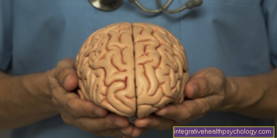
For a variety of reasons, a Imaging your head be inevitable. Using MRI or CTRecordings, doctors can quick statements about possible disease processes fall within your brain. Basically, in every single case decided whether an administration of contrast medium is necessary. Very often, detailed assessments can only be made through contrast enhancement.
E.g. in multiple sclerosis, even the smallest changes in the brain can only be discovered with contrast media. Contrast-enhanced images also provide valuable details in brain tumor diagnostics.
pregnancy
X-ray examinations, i.e. CT and conventional X-rays, are usually not used during pregnancy because radiation is dangerous for the healthy development of the unborn child can be.
With those used in practice MRI machines basically results from research and practice no indication of a possible hazard of the unborn child. Nevertheless, one avoids the investigation as a precaution in the first three months.
Administration of contrast medium should therefore be used during pregnancy extremely cautious be applied. As soon as however potentially life threatening situations occur for the expectant mother and a contrast-enhancing examination could be life-saving, it must Risk of damage to the embryo accepted become.
Please also read our page MRI in pregnancy- is it dangerous?
Conclusion
In summary, are Contrast media It is impossible to imagine modern medicine without it. Both at roentgen, CT as well as MRI, help similar tissue better to differentiate. In the vast majority of cases, the application runs smoothly and those affected feel it hardly any side effects.
Only with some clinical pictures, such as Renal insufficiency or hyperthyroidism, the risks and benefits must be carefully weighed against each other.










.jpg)


