Electroencephalography (EEG)
Synonyms in a broader sense
Electroencephalography, electroencephalogram, (ge) brain wave measurement, measurement of brain waves
English: electroencephalography
Use in medicine
The EEG is an important measure that is often used in examinations in neurology.
Expressiveness
With the help of Electroencephalography (EEG) can basically make statements about the basic electrical activity of the human Brain, via spatially delimited brain activities (Focal findings) and convulsive activities (epilepsy) to be hit. For certain suspected diagnoses (e.g. epilepsy), electroencephalography is absolutely indicated as a diagnostic method. However, an unremarkable electroencephalogram is no guarantee that a specific suspected diagnosis will be excluded. For further clarification, therefore, is often modern imaging procedures (Magnetic resonance imaging of the head , Computed tomography) is used. A "normal" electroencephalogram can, however, provide important information for neurological diagnostics. At brain death (Zero line) the electroencephalogram shows no more electrical activity for at least thirty minutes, which represents an irreversible (irreversible) loss of brain function. You can also use electroencephalography no Statements are made about the personality traits, character or intelligence of the patient.
General
With the help of electroencephalography, the bioelectrical activity of the brain can be registered by means of an electroencephalogram (also known as EEG or brain current image). In neurology, electroencephalography is a diagnostic non-invasive (not penetrating the body) Procedure that is painless and harmless to the patient. It takes between 20-30 minutes to record an electroencephalogram.
Background and preparation for electroencephalography
.jpg)
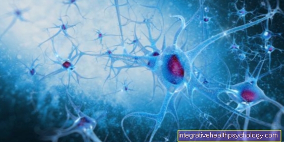
The human brain is constantly activated. This is the case both in the waking and in the sleeping state. This activity is reflected in electrical discharges large aggregates of nerve cells on the surface of the brain, which were generated with electroencephalography (EEG) can be measured using the Head surface (Scalp) at prescribed drainage points Electrodes (thin metal plates) attached, with which the natural voltage fluctuations of the Neurons recorded in the brain. Most electrodes are self-adhesive or are already attached in a suitable cap that the patient puts on over the head (like a swimming cap).
There is usually a contact paste under the electrodes to ensure better contact between the head surface and the electrode. Sometimes it may be necessary to roughen the scalp in certain areas to improve the quality of the dissipation. It is usually done on a routine basis EEG examination a certain number of electrodes are attached to the head surface. The electrodes are on a cable amplifier connected, which receives and amplifies the bioelectrical signals of the brain. These signals are then recorded either on paper or using a computer.
Course of electroencephalography
_2.jpg)
After the electrodes have been attached (approx. 10 min), the patient is asked to remain calm and thereby use the eyes to close or open (Spontaneous EEG). In addition, with certain suspected diagnoses it is advisable to perform an electroencephalogram while awake (Long-term EEG, 24 h) or while sleeping (Sleep EEG) derive.
Sometimes it may be necessary for the patient to be exposed to sensory (visual = stimuli via the eyes, auditory = stimuli via the ears, tactile = stimuli via the skin) or motor stimuli (stimuli via movement) at the same time. This checks the processing of stimuli in the brain (Evoked potentials (EP), event-related potentials).
Electroencephalography is used to determine if epilepsy is suspected Provocation methods (deep breathing, application of bright flashes of light, sleep deprivation, medication). These provocation methods lead e.g. to an increase in the tendency to convulsions, which are characteristic of epilepsy. This is necessary if epilepsy is suspected, as the electroencephalogram between seizures is often normal.
Indications
An EEG examination (Electroencephalography) is carried out especially if the following suspected diagnoses are present:
- Epilepsy epilepsy
- Encephalitis (Inflammation of the brain)
- Metabolic diseases
- Brain damage
- Brain breakdown processes (Creutzfeld-Jakob)
- Sleep disorders / diseases
- Impaired consciousness (coma)
- Brain death
Please also read our page Diagnosis of epilepsy.
Risks, complications, disturbances
Contrary to the widespread assumption that the patient is “energized” during electroencephalography, it must be emphasized that with this procedure only the weak potential fluctuations from nerve cells in the brain flow to the EEG devices, but no current from the device to the electrodes . The electroencephalography procedure is therefore risk-free and side effects are not known.
During the recording of the electroencephalogram, e.g. by heavy sweating or strong movements mean that the electroencephalogram cannot be used.
Helpful for a good derivation of an electroencephalogram (EEG) are also washed hair, as the fat on the scalp can worsen the transfer between the head surface and the electrode.
evaluation
With the help of electroencephalography (EEG) an electroencephalogram is created, on which the course and strength of the bioelectrical activity of the brain are recorded. This electroencephalogram contains waves that are evaluated according to certain frequency patterns (frequency bands), amplitude patterns, local activity patterns and their frequency of occurrence. Generally speaking, it is considered which curves are present, how fast they are, whether they are deformed and whether the curves have certain patterns.
Special computer-aided processes (e.g. spectral analysis) can also be used for evaluation. The frequency bands, which can generally be divided into four categories, are particularly rich in information during the evaluation:
Delta waves
Frequencies from 0.5 to 3 Hz: This frequency band can be observed particularly in deep sleep and is characterized by slow and large amplitudes in the electroencephalogram.
_3.jpg)
Theta waves
Frequency from 4 to 7 Hz: These frequencies occur during deep relaxation or while falling asleep. Slow theta waves are normal in children and adolescents. In the awake adult, the permanent occurrence of theta waves (and also delta waves) is to be assessed as a noticeable finding.
_4.jpg)
Alpha waves
Frequencies between 8 and 13 Hz: These frequencies represent the basic rhythm of the bioloelectric activity of the brain and appear in the electroencephalogram when the patient's eyes are closed and the patient is in a resting state.
_5.jpg)
Beta waves
Frequencies from 14 to 30 Hz: This frequency band shows itself when sensory stimuli occur (i.e. in the normal waking state) or when mental tension.
_6.jpg)


