diaphragm
Synonyms
medical: diaphragm
definition
The diaphragm is a peculiarity of mammals. It is a three to five millimeter thick, dome-shaped, muscular-sinewy plate that separates the chest (thorax) from the abdomen (abdomen) and represents the most important respiratory muscle.
anatomy
Construction:
In terms of tissue technology (histologically), the diaphragm can be divided into two parts. The muscular parts come from the origins of the chest muscles in the lower chest wall. They unite in a central tendon field (Centrum tendineum), which represents the sinewy part.
For orientation, the muscular part of the diaphragm is divided into three sections:
- middle part, behind the sternum = Pars sternalis
- facing the ribs on both sides = Pars costalis
- rear part, facing the back = Pars lumbalis, with right and left thigh (Crus dexter and Crus sinister).
The muscular portion of the Pars sternalis arises from the posterior surface of the xiphoid process (Xiphoid process) and the rectus sheath. The Pars costalis arises from the cartilage of the lower six ribs on the costal arch. The middle portion (crus mediale) of the Pars lumbalis arises from the lumbar vertebrae 1-3 and their intervertebral discs; the lateral part (crus laterale) originates from the second lumbar vertebrae, the 1st tendon arch of the psoasarcade (arcade-shaped tendon strip of the Psoas major muscle, Ligamentum mediale), the 2nd tendon arch of the quadratus arcade (arcade-shaped tendon strip of the quadratus lumborum muscle, ligamentum laterale) and the costal process of the second Lumbar vertebrae to the tip of the 12th rib.
The various muscular parts unite on both sides in the shape of a dome to form a sinewy center and thus form the boundary between the abdomen and chest. Thus it has close neighborly relations with organs of the two body cavities. On the upper side, in the chest cavity, the right lung lobe borders on the right and the left lung lobe and the left Pericardium (Pericard), which is fused with the sinewy center of the diaphragm. On the underside of the abdomen on the right, the liver (with its area nuda fused with the diaphragm) and the right border kidney and on the left the left Liver lobe, the gastric fundus, the spleen and the left kidney to the diaphragm.
Between the muscle parts there are triangular triangles filled with little connective tissue: that Trigonum sternocostale (in the middle, so-called Larey's fissure) in the pars sternalis and the Trigonum lumbocostale (on both sides, so-called Bochdalek triangle).
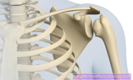
Figure diaphragm

- Diaphragm (red) -
Diaphragm - Chest cavity -
Cavitas thoracis - Abdominal cavity -
Cavitas abdominis - Tendon center of
Diaphragm -
Centrum tendineum - Rib part of the diaphragm -
Pars costalis diaphragmatis - Esophageal slit -
Aortic hiatus - Vena cava hole -
Foramen venae cavae - Aorta in the aortic slit -
Aorta in the aortic hiatus - Loin part of the diaphragm -
Pars lumbalis diaphragmatis - Lungs - Pulmo
The diaphragm separates
the thoracic and abdominal cavities
You can find an overview of all Dr-Gumpert images at: medical illustrations
Passage points of the diaphragm / diaphragmatic gaps:
The diaphragm has openings for conduction pathways and organ connections to pass through. The following overview gives an overview.
Diaphragmatic gap:
Foramen venae cavae (at the level of the 8th thoracic vertebral body-BWK- in the centrum tendineum)
- Passing structures:
- Inferior vena cava
- Ramus phrenicoabdominalis of the right phrenic nerve
Hiatus oesophageus (in the amount of BWK 10)
- Passing structures:
- Esophagus and lying on it
- Trunci vagales anterior and posterior (parasympathetic nerve fibers)
Aortic hiatus (in the amount of BWK 12 / LWK 1)
- Passing structures:
- Descending aorta
- Thoracic duct ("breast milk duct", lymph vessel)
Columns in the medial crus
- Passing structures:
- Right: azygous vein
- Left: hemiazygous vein
- Nervi splanchnici
Fissures between the medial and lateral crus
- Passing structures:
- Sympathetic trunk
Trigonum sternocostale
- Passing structures:
- Internal thoracic artery and superior epigastric vein
Vascular supply of the diaphragm:
Above the diaphragm, the following arteries supply the diaphragm with oxygen-rich blood: arteria pericardiacophrenica dexter and sinister, which arise on both sides of the arteria internal thoracica dexter and sinister, and the arteria musculophrenica dexter and sinister, which continue the course of the arteria thoracica interna. A branch of the thoracic aorta, the superior phrenic artery, also contributes to the supply.
Below the diaphragm, a branch of the abdominal aorta, the inferior phrenic artery, supplies the diaphragm. The venous outflow takes place via veins of the same name.
The diaphragm becomes nervous through the Phrenic nerve from the cervical nerve plexus (plexus cervicalis). He receives parts from the spinal cord segments C3-5. (The "C" stands for cervical, ie located in the neck area). An old motto for this is: "Three, four, five, keeps the diaphragm alive!"
Functional and topographical anatomy of the diaphragm
The relative position of the diaphragm is clinically relevant. The topographical references help with orientation in the chest and with the interpretation of X-ray images. The diaphragmatic domes are clearly visible here. Due to the curvature, a gap in the pleura is lowered between the chest wall and the diaphragm (Pleura) a, the Costodiaphragmatic recess. A further gap arises independently of the diaphragmatic bulge between the ribs and the rear wall of the sternum on the one hand and the anterior pericardial wall on the other. When inhaling deeply, the lungs shift into these reserve gaps. The projection of the diaphragm onto the trunk depends primarily on the breathing position. In the exhalation position, the diaphragm is high up to the 4th rib, with maximum inhalation it can be lowered to the right almost to the 7th rib. The actual position of the diaphragm continues to depend on the type of constitution, age and gender of the person. In addition, it stands because of the asymmetrical position of the Heart left lower than right. Inhaling not only lowers the diaphragm, but also flattens both domes. The extent of movement of the diaphragm can be estimated during inspiration based on the displacement of the palpable edge of the liver. The breathing movement is about six to seven centimeters. When lying down, the diaphragm is higher than when standing due to the pressure of the abdominal organs. Because of the loss of tone, the diaphragm is higher on the corpse than on the living person during exhalation (for the proximity of the organs, see above).
Diaphragmatic breathing

The diaphragm is a huge muscle with a large central tendon attachment. The diaphragm separates the abdomen from the chest (thorax) and is a very important muscle for breathing in general. The diaphragm is particularly important when inhaling (inspiration), as it creates a negative pressure through a muscle contraction and ensures that the abdominal organs are pressed down and thus creates more space for the air flowing into the lungs, so that the lungs can oxygenate red blood cells.
Thus, with every breath, diaphragmatic breathing takes place, which is vital in order to properly fill the lungs and thus the entire body circulation with oxygen and fresh air.
Diaphragmatic breathing is often equated with the term abdominal breathing. In the end, diaphragmatic breathing is all about the fact that the contraction of the diaphragm creates more space for the development of the lungs and allows them to expand a little. Since the diaphragm presses downwards, the abdominal organs are shifted into the abdomen and the abdominal wall bulges slightly. You can understand this with your hands when you place them on your stomach and then consciously inhale and exhale deeply. This is known as abdominal breathing. This form of breathing is caused by diaphragmatic breathing and is therefore often used as a synonym with it.
Diaphragmatic breathing is opposed to chest breathing, in which the diaphragm only contracts minimally and the chest mainly expands upwards to allow the lungs the necessary expansion so that they can absorb the freshly inhaled air.
Diaphragmatic hernia
With the diaphragmatic hernia or the so-called hernia, parts of the abdominal organs shift into the chest cavity through congenital or acquired weak points. This type of fracture is also known as internal hernia, as it is not visible to the doctor from the outside, for example.
A diaphragmatic hernia always occurs at the point of least resistance - "Locus minoris resistentiae". These form the natural penetration points and the muscle-free parts of the diaphragm (Larey Column, Bochdalek Triangle). When the intra-abdominal pressure is increased, abdominal organs can break through into the chest cavity.
The danger here is the entrapment of intestinal loops with the risk of intestinal obstruction. The consequences are severe abdominal pain of unclear cause. A quick operation is indicated here, as otherwise there is a danger to life. With 90% of all cases the hiatus oesophageus is the most frequent fracture and entry portal. Most of the time, the end of the esophagus with the stomach entrance (cardia) "slides" through the hiatus into the chest cavity (axial hiatus hernia or sliding hernia; about 85% of all hiatal hernias). Typical complaints are heartburn, acid regurgitation and a feeling of pressure behind the breastbone after eating, up to nausea, vomiting, shortness of breath and functional heart problems.
Congenital diaphragmatic hernias are found with a relatively high probability of 1: 2000 births in the Bochdalek triangle. The cause is the incomplete closure of the diaphragm during embryonic development. Abdominal viscera pass through here and press on the heart and lungs. In this case, too, rapid surgery is necessary due to the acute danger to life.
Antenatal ultrasound diagnostics can show the defect. The localization is almost always on the left. The reason is simple: the liver is on the right. A hernia in the trigonum sternocostale, i.e. behind the breastbone (morgagnia, parasternal hernia), is possible, but is less common.
Read more about diaphragmatic hernia.
Pain in the diaphragm
The diaphragm (Diaphragm) is ours most important auxiliary respiratory muscle and supported especially inhalation (inspiration), since exhalation (Expiration) largely passive runs out and does not require any muscle support.
Since the diaphragm also has the belly (Abdomen) with the entire digestive tract (gastrointestinal tract) of ours Rib cage (Thorax) separates, pain in the area of the diaphragm must always be taken seriously. Most of time the pain is harmless causes.
Inexperienced singers can your diaphragm through a Improper loading have overloaded, as the muscles of the diaphragm are used especially when singing. Also prolonged fits of laughter can possibly lead to pain in the diaphragm the next day. The pain should only be briefly occur and rather than uncomfortable really felt to be extremely painful.
At stronger pain in the diaphragm it can be, among other things, a Inflammation of the diaphragm act. While this condition is extremely rare, it can occur because of a infection, as a result of a (partial) Displacement of the stomach in the rib cage and the resulting Exit of Stomach acid in the area of the diaphragm or due to psychological stress arise.
In the latter case, they are usually the annoythat supply the diaphragm, affected and lead to a strong feeling of pain in the area of the diaphragm.
Is the Inflammation of the diaphragm infectious conditionally, it usually comes next to the Pain in the diaphragm with accompanying Shortness of breath and problems laughing and walking, too fever, Body aches and general malaise. Since the shortness of breath makes the patient very vulnerable, it is the Inflammation of the diaphragm in the late stage by one life threatening illness. In order to contain the infection, it is usually advisable to give Antibiotics.
If the pain is very severe and it restricts breathing, the patient should also Pain medication take in.
In addition to the Inflammation of the diaphragm however, it can also be a Diaphragmatic hernia cause pain in the diaphragm. A diaphragmatic hernia is a weak spot in the field of Muscles or Tendons of the diaphragm through which the Digestive organs (mostly esophagus or intestines) in the chest be relocated. This can lead to severe pain, shortness of breath, malaise, nausea and vomiting. It occurs especially in patients in whom the esophagus and stomach slide further and further into the chest area heartburn and more often nausea. In most cases, a diaphragmatic hernia does not have to be treated; it is only recommended if the problems become too severe Heartburn medication (Antacids, Proton pump inhibitors etc) or in case of doubt also one surgery.
You can find more information about this on our website Pain in the diaphragm.
Diaphragmatic cramp
A diaphragmatic spasm is one that starts suddenly Contract of the diaphragm, which extends through hiccup and can express severe pain in the upper abdomen. Possible causes include a diaphragmatic hernia or nerve irritation.
Pinched diaphragm

The diaphragm provides crucial support for people when breathing in and, as a large, muscular and sinewy plate, separates the chest from the abdominal area with the entire digestive tract.
If the diaphragm is pinched, it is a diaphragmatic hernia or diaphragmatic hernia. Due to a weak point in the area of the muscles and the tendons of the diaphragm, parts of the gastrointestinal tract (usually the esophagus, possibly also parts of the stomach) shift through the diaphragm up into the chest. The diaphragm is only pinched indirectly. Actually, it is more a question of an expansion of a physiological opening in the diaphragm. Since the esophagus connects the mouth with the stomach and lies in the chest, it has to pass through the diaphragm. Therefore the diaphragm is not a continuous plate, but has holes; The esophagus passes through one of these openings (hiatus oesophageus) and opens into the stomach.
If it comes due to increased pressure in the area of the abdomen (abdomen), so that the hole through which the esophagus passes is widened, it is possible that the upper part of the stomach (in rare cases also parts of the intestine) enters the chest area slide and cause problems.
The diaphragm is only indirectly pinched, but it can feel that way to the patient. Often there is also heartburn, nausea and sometimes even vomiting and pain in the area of the diaphragm. In this case, the patient should either take heartburn medication (such as antacids or proton pump inhibitors) or consider surgery.
Tense diaphragm
The diaphragm is the most important Auxiliary respiratory muscles of humans and makes sure that we breathe in (to inspire) can. Exhalation (expiration) takes place by itself in resting breathing, but when we Sports operate and us physically exerting yourself, the diaphragm must also die Support exhalation. But not only with the breathing the diaphragm has a crucial role. Also at Speak, but especially with Scream or at To sing, the diaphragm is extremely important.
Since the diaphragm is a big muscle With a sinewy plate in the middle, it can also be in the area of the diaphragm Tension come. The diaphragm becomes tense especially when we do it overload. On the one hand, this can be the case with an enormous amount of sporting activity, if the patient had to inhale and exhale a lot and overloaded the diaphragm as a result; on the other hand, it can be that the diaphragm becomes tense after a loud argument or longer singing, similar to one aching.
Especially inexperienced singers can through the incorrect use of the diaphragm ensure that the diaphragm is tense. Here it is advisable to seek out a suitable singing teacher to prophylactic exercises to get shown.
It is general harmlesswhen the diaphragm is tense and the patient should especially prevent it the next time suitable breathing training do and join often tense diaphragm advice from one Singing teacher, Voice trainer or one Physiotherapists search.
Exercise the diaphragm
The diaphragm (Diaphragm) is needed when speaking, especially when we are speaking louder or shouting. The diaphragm is also very important for Singer and singers but also for flute players or in general for Wind players. These use the diaphragm constantly in the form of Abdominal breathing and therefore need a well-trained diaphragm.
In order to train the diaphragm, it is therefore most important to train abdominal breathing. Especially in the Forget stress many patients on a steady and calm dto pay attention to instead just being breathed into the chest. However, it is important to exercise abdominal breathing and thus the diaphragm, as this is on the one hand Constipation (Constipation) prevent and on the other hand too Diaphragmatic hernias (Hernias) prevent can.
A very good exercise is to yourself lay flat on your backto yourself completely relax and the Hands on stomach to lay. Now you should go deep into your stomach inhale and exhale. This can be done using the handslying on their stomach check. At the Exhale the belly should be something flatter be at breathe in the belly should be outward bulge out.
In order to be able to train the diaphragm in the office without lying flat on the floor in front of your colleague, you can also use the To sit, at straight back and head straight the hands lie on your stomach and try again to breathe into your stomach while doing the Follow breathing movements. These two exercises should help to train the diaphragm and are especially important for musicians and singers.
Inflammation of the diaphragm
By a infection, but also by high mental stressinflammation of the diaphragm (Diaphragmatic disease) come. In general, a diaphragmatic inflammation is extremely rare, however, as it is associated with severe Pain and consequently also strong restrictions connected, it should not be underestimated.
It is important to remember above all that a diaphragmatic inflammation rarely as a primary disease occurs, but that mostly a infection or Digestive tract discomfort (Stomach and intestines). Especially with the well-known heartburn (Reflux esophagitis) can be caused by the acidic stomach acidthat possibly Contact with the diaphragm gets to one Diaphragmatic irritation come. Because of this irritation, the diaphragm is very much lighter by infectious agents vulnerable and thus it can lead to diaphragm inflammation more quickly.
Another, but extremely rare, cause is that Infestation with trichinae. This is Roundwormsthat enter the body through the consumption of raw meat, for example, via Bloodstream come to the area of the diaphragm and there one Inflammation of the diaphragm can trigger.
The symptoms of diaphragm inflammation are characteristically always associated with severe diaphragmatic pain. Since the diaphragm especially in the inhalation (Inspiration) is needed is this particularly painful. In addition, laughing, coughing or talking loudly can lead to greater pain, so that the patient on the one hand breathe more shallowly and on the other hand often one Relieving posture take (bent slightly forward) to relieve the diaphragm. In addition to the pain, it can also cause a parasite infection Rise in fever come.
A diaphragmatic inflammation can be over imaging procedures on the other hand, there is the possibility of the patient Draw bloodto the type of Pathogens, as well as that extent the diaphragmatic infection to determine. Depending on the cause of the infection, you can get specific Medication (anti-infectives) give and if necessary Pain medicationto allow the patient again breathe symptom-free can and again without any problems laugh and especially to cough can, as the latter is a reflex that may be vital in some circumstances.
Here you can find further information on the topic: Inflammation of the diaphragm
Summary
The diaphragm fulfills vital functions in our body. It not only separates the chest from the abdomen and thus contains important passage areas for structures from the chest into the abdomen and / or vice versa, it is also the most important respiratory muscle, which in the event of failure usually has fatal consequences for the patient concerned.


.jpg)