ankylosing spondylitis
Synonyms in a broader sense
Ankylosing spondylitis (AS), ankylosing spondylitis, spondylarthropathy
Rheumatism, Rheumatoid arthritis, Psoriatic arthritis, methotrexate
English: Ankylosing spondylitis
definition
Bechterew's disease is one of the most common inflammatory rheumatic diseases. It belongs to the group of so-called spondyloarthropathies, to which i.a. also include psoriatic arthritis, inflammatory bowel disease arthritis, Lyme arthritis (borreliosis), rheumatoid fever and reactive post-streptococcal arthritis.

The inflammatory changes are mainly found in the area of the Spine and to the Sacral and iliac joints (SI joint). In 20-50% of patients there are others as the disease progresses Joints (e.g. hip joint and knee joint).
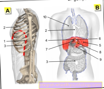
20% of patients also suffer from inflammation from:
- Tendon attachments (enthesopathy)
- eye
- Intestines
- heart
- kidney and
- lung.
history
The disease was first discovered in 1884 by Adolf Strümpell from Leipzig on the basis of two patients with a complete stiffening of the spine and joints.
More reports followed from Vladimir von Bechterew (1886-1927) from St. Petersburg and from Pierre Marie from Paris.
root cause
The cause of Bechterew's disease is unknown. The association of the disease with genetic characteristics is known, in particular with the human leukocyte antigen HLA-B27. Over 90% of those affected are HLA-B27 positive. In Germany, around 8% of the population are HLA-B27 positive, of which 2-5% develop Mb. Bechterew, i.e. over 90% of HLA-B27 positive people stay healthy.
In the case of first-degree relatives suffering from illness, the risk of developing Bechterew's disease is 20%, and 60% in the case of identical twins. In Germany there are about 800,000 patients with Bechterew's disease.
As with other inflammatory rheumatic diseases, certain bacterial infections are discussed as triggers.
At the onset of the disease, the average age of the patients is 26. Men are two to three times more likely to be affected than women.
Symptoms / complaints
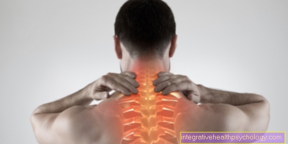
In about 75% of the patients, deep-seated lower back pain is the first symptom. The onset is usually gradual and is before the age of 40. Persistent are characteristic complaints over three months, occurrence of the complaints especially in the second half of the night, in the morning and after a long break. Typically, symptoms improve with exercise and respond well to nonsteroidal anti-inflammatory drugs (NSAIDs). In addition to the sacrum and iliac joints, the transition from the Thoracic spine to Lumbar spine (Th8-L2) affected.
In the course of the disease, the mobility of the spine is increasingly restricted, up to complete stiffening. In extreme cases, in the late stage, the patient can no longer raise the line of sight above the horizontal by blocking the thoracic vertebrae in a bent position and stretching the cervical spine.
By Participation of the Rib-vertebral joints breathing movement can be restricted. Pain in the anterior chest wall can be caused by inflammatory changes in the sternum-clavicular joints (sternoclavicular joints), the Sternum (Synochondrosis manubrio-sternalis) as well as on the costal cartilage (enthesitis).
In 20% of patients, the disease appears for the first time in the form of joint inflammation (arthritis) of a peripheral joint, typically in one or a few joints (mon- or oligoarthritis) in the area of the legs.
The inflammatory changes also lead to changes in the tendon attachments. Because of the particular stress and prominence, these appear in about 20% of patients in the form of a heel pain, sometimes also in the area of the large rolling hillocks (greater trochanter), on the ischium or on the ischium Iliac crest.
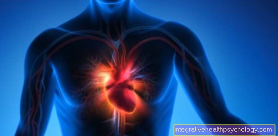
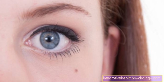
Outside the musculoskeletal system, Bechterew's disease can also become symptomatic as an inflammation of the eye (iridocyclitis). Acute onset of pain in one eye, sensitivity to light and impairment of visual acuity occur. Further manifestations can be in the area of the heart and blood vessels in the form of a Aortic regurgitation and Cardiac arrhythmias and occur in the area of the intestine in the form of ileitis or colitis. Involvement of the lung (bilateral apical pulmonary fibrosis) and the kidney (IgA nephropathy).
A complication after a long-term course with high inflammatory activity can be a so-called. Amyloidosis (Deposition of proteins in internal organs with subsequent disruption of organ function). Another complication in the later stages of the disease is the increased risk of bone fractures, especially in the area of the spine. Due to the stiffening, bone fractures can occur even with minor trauma, as the bone has lost its elasticity.
diagnosis
The diagnosis is made on the basis of symptoms and physical examination in conjunction with laboratory and X-ray findings. Internationally enforced for diagnosing Morbui. Bechterew have the modified ones New York criteria from 1984:
- diagnosis
1. Clinical criteria:
2. Radiological criterion- Deep lower back pain and stiffness for more than 3 months. Get better with exercise, but not with rest.
- Limited mobility of the lumbar spine in the sagittal and frontal planes.
- Restricted breathing excursion of the chest to <2.5 cm (corrected for age and gender).
- Inflammatory changes in the area of the sacrum and iliac joints (sacroiliitis> grade 2 on both sides or grade 3-4 on one side).
- graduation
1. Safe ankylosing spondylitis:
if at least one clinical criterion and the radiological criterion are met.
2. Probable Mb. Bechterew:- When three clinical criteria are met.
- If only the radiological criterion without clinical criteria is met.
The problem is that certain x-ray changes only appear after an average of 5-9 years. Manifestations outside the joints are common and can be the first manifestation, so the early forms of ankylosing spondylitis often cannot be diagnosed.
Laboratory values
Approx. 90% of patients with ankylosing spondylitis have the human leukocyte antigen B27 in their blood. However, the HLA-B27 determination is not suitable as a search test. However, if ankylosing spondylitis is suspected based on the symptoms, physical examination and radiological findings, a positive HLA-B27 increases the probability of the diagnosis.
Increased inflammation levels in the blood, such as CRP (C-reactive protein) and an accelerated sedimentation rate (ESR), correlate with the inflammatory activity and can therefore be used to monitor the progression. In the case of mild courses, however, these can also be in the normal range.
Read more on the topic: Levels of inflammation in the blood
As part of the diagnosis of Bechterew's disease, HLA is also determined. For more information, read the following article: HLA - Human Leukocyte Antigen
roentgen
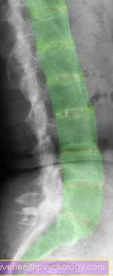
The radiological changes in the area of the sacrum and iliac joints are of great importance for the diagnosis of ankylosing spondylitis, but are not suitable for assessing the course of the disease Sacroiliitis (Inflammation of the sacrum and iliac joint) are a blurred joint contour with near-joint sclerosis (compression of the bone) and erosions (bone dissolution) (so-called colored picture).
In the area of the spine and peripheral joints, the radiological changes are more suitable for assessing the progression. They are the result of inflammatory destruction and mostly frustrating reparation processes.
The following are observed on the X-ray:
- Erosions
- Sclerotherapy
- Joint space narrowing
- blurred joint contours
- Syndesmophytes (calcification of the ligaments of the spine, so that the vertebral bodies are functionally connected) and
- Bone spurs.
Spondylodiscitis, box vertebra formation, spondylophyte formation, bony bridging and ultimately complete joint or vertebral body fusions (so-called bamboo sticks) occur on the spine.
Magnetic resonance imaging (MRI) of the pelvis and spine
Much earlier than in X-ray image are the inflammatory changes in the sacrum and iliac joints (SIJ) and the spine with the Magnetic resonance imaging (MRI) to represent.
The MRT can also be used to make a statement about the intensity of the inflammation, so that the method is also suitable for assessing the progress and monitoring the success of the therapy.
However, it is not possible to depict all regions affected by ankylosing spondylitis with the same quality with an MRI. Therefore, there is an income for the ISGs MRI of the pelvis or the LWS with ISG in question.
If you want to be able to assess your entire spine MRI of the spine be driven.
Sonography / ultrasound
As a cost-effective and side-effect-free method, sonography is used to record and monitor the course of peripheral joint inflammation and inflammation of the tendon insertions. It can also easily be carried out as a dynamic examination and in side-by-side comparison.
General information on the topic can be found at: Sonography
Summary
Bechterew's disease is one systemic inflammatory disease unknown cause from the group of spondyloarthropathies.
The main places of manifestation are the sacrum and iliac joints (SI joint), the transition from the Thoracic spine to Lumbar spine and with peripheral joint involvement of hip joint and knee joint. Often there is also inflammation of the tendon attachments and involvement of the eye (iridocyclitis).
They typically occur persistent Pain and an increasing restriction of movement.
The diagnosis is made clinically (by examining the patient) and radiologically (by X-rays, MRI; CT, Scintigraphy Etc.).
Laboratory values can be a positive HLA-B27 or increased inflammation values confirm the diagnosis. In order to contain the inflammatory process and the progressive stiffening or destruction of the joints, forced therapy must be initiated at an early stage. Physiotherapy / physiotherapy and drug therapy. If conservative therapy measures fail, surgical therapy measures are used.


.jpg)