Empyema
Synonyms
Collection of pus, pus cavity
definition
If pus accumulates in a prefabricated body cavity in the course of an inflammation, the specialist calls this accumulation empyema.

General
As part of an inflammatory reaction, especially in the case of bacterial infections, pus often develops.
Pus is usually yellow and viscous, but generally quite variable in its nature and composition.
Figuratively speaking, pus is what remains of an immune defense battle: dead pathogens (mostly bacteria), dead defenders (leukocytes) as well as tissue waste resulting from collateral damage. This is caused, for example, by pathogen toxins or the body's own substances (such as so-called proteases, which cut up proteins) that damage the surrounding body tissue.
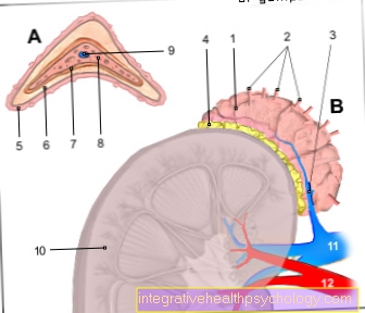
If the inflammatory process takes place very close to or even in an already existing body cavity, it is obvious that the pus that develops collects in this cavity: an empyema develops. Depending on which body cavity is affected, empyema can be described in more detail.
Empyema can also develop in the pleural cavity, i.e. between the lung envelopes, and have undesirable consequences. At this point you should also deal with the topic: Pleural Empyema - What's Behind It?
causes
Primarily pus-forming germs (pyogenic pathogens) are responsible for purulent inflammations. Many bacteria of the groups of staphylococci and streptococci are pus-forming germs and cause the majority of the clinically observed empyema.
In exceptional cases, viral infections, diseases caused by parasites and autoimmune diseases can also take a purulent course.
It is extremely unlikely that the pus formation is so pronounced in terms of quantity that empyema can develop.
A gallbladder empyema usually develops in the context of a gallbladder inflammation - mostly originally triggered by a gallstone - in which bacteria have colonized it.
Pleural empyema, i.e. a collection of pus in the gap between the lungs and the chest wall, is often caused by inflammation of the lung (pleurisy). This is a common complication of bacterial pneumonia, such as pneumococci. Another complication can be that pus can simply build up in the lungs.
Empyema in the maxillary sinuses, in the abdominal cavity (purulent peritonitis) or in joints (pyarthrosis) are also possible and are also usually an expression of an underlying bacterial infection
Symptoms and consequences
In addition to general symptoms from an underlying infection such as fatigue, fever etc., are due to the local inflammatory reaction too pain, Swelling, redness, warming and Functional restrictions possible. The severity of these symptoms depends on the location and extent of the empyema.
Since the purulent interior of an empyema is suitable for immune cells (Leukocytes) and is difficult to access for medication, empyema can sometimes persist for a long time without medical intervention. The pus can also serve as a food basis for other germs.
Furthermore, every empyema harbors the risk of germs from the accumulation of pus into the Bloodstream and can settle in another place (scatter) or the clinical picture of a life-threatening Blood poisoning Trigger (sepsis).
Ultimately, in the course of the healing of a (purulent) inflammation, adhesions can occur, which can subsequently cause problems, especially in the abdomen and in the gap between the lungs and the chest wall.
diagnosis
Sometimes it can be done by a conscientious medical interview (Anamnesis) and physical examination an urgent suspicion of empyema and its location can be made.
Also a laboratory one Examination of the blood can continue through an existing increase in so-called inflammation parameters.
However, the best way to localize an empyema is through the use of imaging techniques such as Ultrasonic (Sonography), MRI or CT.
Theoretically, both the presence and the location of an empyema can be reliably determined intraoperatively.
therapy

The inside of a pus cluster is generally for Immune cells and Medication (such as useful here Antibiotics) hard to reach.
From a certain size of an empyema, a systemic therapy with a therapy adapted to the pathogen (and its resistances) is necessary Antibiotic therapy (in tablet form or via an indwelling cannula) to a operational opening to think of the cave to drain the pus.
In some cases it can also be useful to rinse the body cavity not only with disinfecting solutions, but even with it Antibiotic solution or temporarily a little chain, sponge, etc. bring in.
Is it a collection of pus in the gap between lung and Chest wall (Pleural space) or in the Abdominal cavityWhere there is a risk of sticking, a special solution can be used to prevent this.
The procedure is usually concluded with the inlay of a drainage, which can still suck for a discharge of secretion and can thus prevent an immediate new formation of the empyema. This drainage then remains Hours to days lie; can even require multiple exchanges in extreme cases.
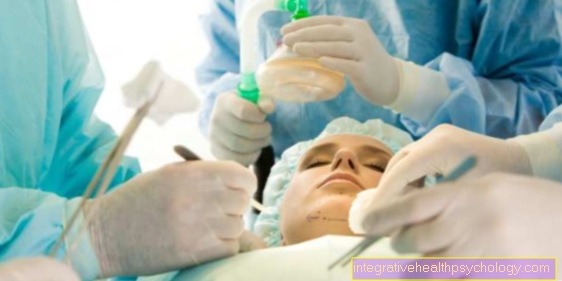
Depending on the case and location, the procedure is carried out either under local anesthesia or under general anesthesia in the operating room.
forecast
In principle, empyemas can be treated well. Whether there are complications such as a Blood poisoning or stop sticking after healing, depends above all on whether intervention was carried out early enough and correctly.
It should be noted, however, that empyema is only an expression of a disease. Whether, and if so, how quickly a cure is possible, depends above all on the underlying disease, which secondary diseases are present, and the general condition and age of the patient.
prophylaxis
If an operation has been carried out in or near body cavities, an artificial drain is created from this cavity in the form of a drainage conceivable as a prophylaxis against the formation of empyema.
Without such a drain, there could be an accumulation Wound secretion serve as a breeding ground for bacteria and thus as a basis for the formation of empyema.
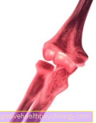
Empyema in the knee
Empyema is an infection of the soft tissues. This is different from the abscess Accumulation of pus in pre-existing body cavities characteristic of empyema. Joint empyemes represent one of the empyemes emergency because, if left untreated, they become a Destruction of the joint can lead within a short time.
The knee joint represents such a body cavity. Knee joint empyema can be called Result of injuries, Broken bones or Operations arise. The pathogens reach the joint directly through open wounds and open bone fractures, but they can also pass through the Blood circulation from other parts of the body be forwarded to the knee.
Also medical measures like a Reflection of the knee joint or injections into the joint as well as open operations on the knee unfortunately promote the penetration of bacteria, although they are carried out under conditions that are as germ-free as possible.
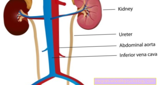
Certain Pre-existing illness such as cancer, diabetes mellitus, gout, liver and kidney diseases or infections such as HIV, immune deficiencies and diseases of the blood vessels such as peripheral arterial disease increase the risk of developing empyema in the knee. This leads to a purulent, painful effusion, which must be opened and cleaned immediately.
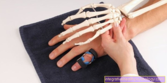
One is typical Restriction of movement, swelling, Redness and overheating of the affected knee. Furthermore can also fever occur.
The basic measures are cooling and immobilization and elevation of the knee. Furthermore, pain medication and thrombosis prophylaxis are administered before the Joint opened and cleaned becomes. In addition, antibiotics are given against the pathogens.
Empyema in the maxillary sinus
Also the Maxillary sinus can from one Empyema to be affected. The maxillary sinus (sinus maxilaris) is one of the Sinuses.
When there is inflammation, one speaks of one Sinus infection (Sinusitis). There can be various reasons for this.
A collection of pus in the maxillary sinus is called maxillary sinus empyema. This can be used as a Complications of chronic sinus infection or caused by the entry of pathogens in other ways. Broken bones in the midface and upper jaw allow germs to enter the maxillary sinus. Inflammation of the upper teeth can be carried into the maxillary sinus.
Are typical Pain when buying, Toothache, Fever and a facial pain. Since there is a risk of the infection spreading to the meninges or the development of a venous thrombosis, quick action must also be taken here. The therapy includes antibiotics and an endoscopic removal of the inflamed and purulent mucous membrane, as well as cleaning the maxillary sinus.