Inflammation of the ethmoid cells
introduction
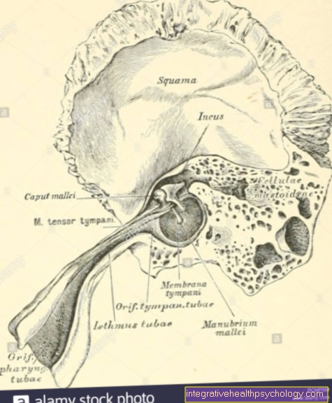
Ethmoid cells (lat. Ethmoid sinus, also cellulae ethmoidales) are a number of air-filled spaces in the ethmoid bone (ethmoid bone). A distinction is made between anterior and posterior ethmoid cells, which form the ethmoid labyrinth. Together with the maxillary, sphenoid and frontal sinuses, the ethmoid cells belong to the paranasal sinuses. As such, these can also become inflamed and cause pain.
Read more about the topic here: Ethmoid cells

As with all paranasal sinuses, the main function of the ethmoid cells is presumably to be found in the weight savings (pneumatization of the bone) resulting from the cavity with air filling. Other functions are still being researched and are considered controversial.
causes
Due to the connection of the ethmoid cells to the outside, infections that originally develop in the nose area can migrate to the paranasal sinuses, i.e. also to the ethmoid cells. One then speaks of a sinus infection, a sinusitis. Mostly these are inflammations caused by viral pathogensthat have already caused discomfort in the main nasal cavity. But also bacteria can be the cause of an inflammation of the ethmoid cells or can be secondary to an already weakened area. While in adults the maxillary sinus is often the site of a sinus infection, it hits in children most likely the ethmoid cells. Often it comes to one Accumulation of secretion and pus inside the cavities, because the inflow and outflow path is only a relatively narrow gap.
Symptoms
Such an inflammation of the ethmoid cells is noticeable with:
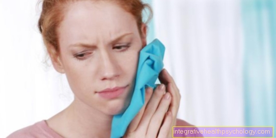
- Sensation of pressure over the forehead and nose as well as below or behind the eyes
- a headache
- a previous or still current cold (rhinitis)
- occasionally fever
Most of the time, the symptoms increase when bending and leaning forward.
You might also be interested in this topic: Inflammation of the sinuses
diagnosis
These typical Symptoms are usually sufficient to make a diagnosis of sinusitis. Especially in the case of severe unclear courses or unclear localization, there is an additional one Nasoscopy into consideration. With this, the doctor looks at the nasal spaces from the inside with the help of a rhinoscope and can thus assess the condition of the mucous membranes. In addition, both a X-rays as well as computed tomographic images the nose and sinuses.
therapy
Acute viral sinusitis usually heals completely within a few days to weeks. That is therapeutic Use of decongestant drugs useful as well as the Taking painkillers and, if necessary, antipyretic preparations.
The same applies to acute bacterial infections that occur for the first time. If there is a suspicion of a bacterial cause of the disease, a antibiotic indexed. Also Cortisone based nasal sprays can fight the inflammation locally.
In some cases it happens long lasting coursesthat can become chronic and then break out again and again (recurrent chronic sinusitis). In the event of unsuccessful attempts at therapy or above-average infections, the next step in the therapy concept is a surgical rehabilitation of the entire paranasal sinuses to disposal. This is usually done endoscopically through the nose (transnasal access) so that no large incisions are necessary. In the course of the operation, pus and excess secretion are removed, all sinuses are rinsed and any anatomical peculiarities that can promote inflammation are eliminated. These include, for example, benign mucosal growths (polyps) or a crooked nasal septum. Part of the frequently inflamed mucous membrane can be removed, thus reducing the future risk of infection.
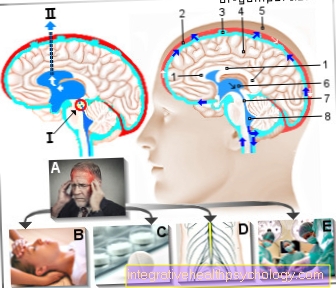
forecast
Inflammation of the ethmoid cells, which often affects small children, usually heals quickly and without complications. In very rare cases, it can spread to neighboring organs, for example the eye socket or the meninges or the brain. Without treatment, there is also the risk of periosteum inflammation.