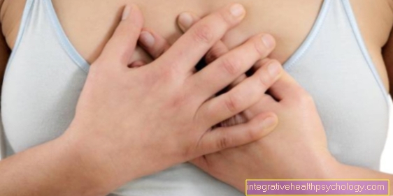
Diseases of the aorta
The most common diseases of the aorta
Find out more about the following diseases of the aorta:
- Atherosclerosis
- Aortic aneurysm
- Aortic dissection
- Coarctation of the aorta
- Marfan's Syndrome
- Aortic arch syndrome
- Takayasu arteritis
- Aortic rupture
- Aortic stenosis
- Aortic regurgitation

Aortic aneurysm
An aortic aneurysm is a congenital or acquired vascular wall sac. A real aneurysm affects all layers of the wall. In contrast, in the case of a false aneurysm, only the intima and the media are affected, the adventitia remains intact.
Causes for such a bulging can be:
- Atherosclerosis
- Infections
- Inflammation
- Trauma (accidents)
- congenital genetic connective tissue diseases such as Marfan syndrome
It is not uncommon for one to appear Aortic aneurysm, especially in the abdominal area (Abdominal aortic aneurysm), without symptoms and is discovered more by chance. If the vessel wall tears, there is an acute risk of death for the person affected by bleeding to death. A rupture can also occur without an aneurysm in accidents (e.g. car accident with impact on the steering wheel).
You can find out more about this topic under our topic: Aortic aneurysm.
Aortic rupture
The aortic rupture describes a rupture of the aorta, usually caused by an accident or trauma, which is life-threatening for the patient and must be treated surgically as soon as possible.
A typical cause is a car accident with a so-called deceleration trauma. This describes the interruption of a jerky movement, as occurs in the event of a crash in a car. The rupture of the aorta is often found in the isthmus area, i.e. the transition from the aortic arch to the section of the aorta in the chest. The consequences are extreme pain and severe blood loss, which leads to shock.
An X-ray or CT image can be made for diagnostic purposes. If the suspicion is confirmed, an operation must be initiated as soon as possible and the aorta sutured again at the ruptured site. Unfortunately, the blood loss is often so pronounced that the patients die relatively quickly after the aortic rupture and treatment in the hospital is too late.
Read more on the subject at: Aortic rupture
Aortic stenosis
Both the primary narrowing of the aorta and the aortic valve stenosis are referred to as aortic stenosis. Aortic stenosis is often caused by arteriosclerosis, with the deposition of fat, connective tissue, thrombin and calcification of the wall layer of the vessel and the lumen of the artery becoming smaller over time. The risk of arteriosclerosis of the aorta arises from the increased mechanical stress on the constricted vessel, which can lead to a tear in the vascular wall with bleeding and / or the formation of a thrombus (vascular occlusion).
Unfortunately, the aortic stenosis caused by atherosclerosis goes unnoticed for a long time. Signs such as dizziness, tightness in the chest or motor or neurological deficits during even light physical activity can indicate massive arteriosclerosis and require a medical clarification. Atherosclerosis, however, is age-dependent and almost always present in people over 80 years of age. However, aortic stenosis can also be caused by congenital malformations of vessels, but this rarely occurs.
Read more on the topic: Aortic stenosis
Aortic stenosis
Aortic valve stenosis is a disease of the heart in which the aortic valve becomes narrowed. In medicine it is often referred to as aortic stenosis. The causes of aortic valve stenosis vary with age. The valve is most likely to have calcification in the elderly. If the stenosis occurs in younger people, the cause is usually a congenital disorder of the valve. Furthermore, rheumatic fever can lead to aortic valve stenosis.
Due to the narrowing of the aortic valve, the heart has to pump against stronger pressure in order to transport the blood from the heart into the body, which causes the heart muscle to become thicker and thicker (hypertrophied) and becomes weaker in the long term and is no longer able to supply the body with sufficient blood (Heart failure). This manifests itself primarily in the form of shortness of breath, dizziness and angina pectoris. If these symptoms occur, the risk of dying is greatly increased, as the heart is already severely affected by the difficult pumping. Therefore, the therapeutic focus is then on the operation in which the valve should be replaced in order to prevent further consequential damage.
Read more on the subject at: Aortic stenosis
Coarctation of the aorta
Coarctation of the aorta describes a naturally occurring narrow point located on the aortic arch between the left subclavian artery (Sinsitra subclavian artery) and the ductus arteriosus, a connection in the bloodstream that dominates prenatal development. This connection is closed after the birth, leaving a natural (physiological) Narrowing. If this is very pronounced, a pathological coarctation of the aorta (narrowing) can occur, which is often congenital and can lead to significant circulatory disorders with organ damage.
Further information is also available under our topic: Coarctation of the aorta
Atherosclerosis
Atherosclerosis describes the calcification of blood vessels, which can lead to changes in blood flow.
It is not uncommon for the aorta to be affected. Blood components, connective tissue, fats or even lime are deposited in the vessel walls. The disease develops over a long period of time and is often the result of malnutrition with:
- too high blood lipids
- lack of exercise
- obesity
- high blood pressure
- Diabetes
- Smoke
These changes cause the elasticity to decrease, which means that the vessel becomes stiffer, which impairs the function of the air chamber. The vessel wall can also narrow (stenosis) or widen (aortic aneurysm). However, the plaques that have formed can also tear open and cause blood clots (thrombi) to form. Such thrombi can narrow the vessel so that the blood flow is impaired or even loosen and get stuck in smaller vessels, which can lead to an infarction of the heart, brain or abdominal organs.
Read more on this topic: Calcifications in the abdominal artery
Aortic regurgitation
Aortic valve insufficiency describes the inability of the aortic valve to close completely, creating a kind of small leak. As a result, the pressure that has to be built up in the heart in order to effectively pump the blood into the body can only be built up to a reduced extent. The whole thing hardly expresses itself at first, but in the long term it leads to insufficiency of the heart muscle, as it tries to compensate for the aortic valve insufficiency and is not permanently aligned with it.
The causes can be acute in the form of an infection (mostly bacterial) or chronic in the form of congenital damage to the aortic valve. The decisive factor in therapy is whether symptoms are already apparent. If this is not yet the case, the heart failure can be treated with medication. If the person concerned shows signs of chronic heart failure with, for example, increased fainting spells (Syncope), an attempt should be made surgically to reconstruct or replace the aortic valve, depending on the extent of the valve insufficiency.
Read more on the subject at: Aortic regurgitation
Aortic arch syndrome
Aortic arch syndrome is a narrowing of several or all of the branches of the aortic arch. The aortic arch itself can also be narrowed (stenosed) be. The main cause is vascular calcification. Sometimes an autoimmune disease (Takayasu arteritis) is also found to be the cause.
The symptoms depend on the degree and location of the narrowing. There may be discomfort and pain in the arms. The arms are also often cool and pale, the pulse cannot be felt or is only weakly palpable. A large difference in blood pressure between the two arms can be an indication of this, depending on where the stenosis is.
If there is insufficient supply to the brain, symptoms similar to attacks, such as:
- Speech disorders
- dizziness
- or visual disturbances.
Takayasu arteritis
This Takayasu arteritis, named after the person who first described it, is a systemic vascular disease that primarily affects the large arteries of the elastic type. The formation of granulomas in the media leads to scarring (internal scars). The first symptoms, however, are first to be found in the muscles and joints, accompanied by the so-called B symptoms (night sweats, fever, weight loss). It is only in the course of the disease that there are narrowing and occlusion of the vessels.
Marfan's Syndrome
In Marfan's syndrome there is a weak connective tissue due to a genetic mutation of chromosome 15. This connective tissue weakness, along with numerous other symptoms, often leads to complaints such as heart valve defects or aortic dissections.
You can find out more about this under our topic: Marfan's Syndrome
Illustration of the aorta

- Ascending aorta -
Pars ascendens aortae - Aortic arch - Arcus aortae
- Thoracic aorta
(descending aorta) -
Thoracic aorta - Aortic slit of the diaphragm -
Aortic hiatus - Abdominal aorta
(descending aorta) -
Abdominal aorta - Aortic fork - Aortic bifurcation
- Trunk of the liver, spleen and ma
gene arteries - Celiac trunk - Humerus artery -
Brachial artery - Common pelvic artery -
Common iliac artery - External head artery -
External carotid artery - Cervical artery (common head artery) -
Common carotid artery - Clavicle artery -
Subclavian artery - Axillary artery - Axillary artery
- Diaphragm - Diaphragm
- Renal artery - Renal artery
- Radial artery - Radial artery
- Ulnar artery - Ulnar artery
You can find an overview of all Dr-Gumpert images at: medical illustrations