Hemothorax
definition
The hemothorax describes one Accumulation of blood in the patient's chest cavity. It represents a special form of pleural effusion Pleural effusion it is an accumulation of fluid between the lungs and the pleura, the two so-called Pleural leaves. Together they make that Pleura ('Pleura"). This effusion can have different causes and different compositions.

A hemothorax occurs when the fluid in the pleural effusion contains at least half as many solid blood components as the blood. Often a hemothorax occurs combined with a Pneumothorax which is an accumulation of air in the pleural space and is usually caused by an injury to the lungs.
causes
Through a accident or the like, there may be external force / force on the chest. Here you can Vessels of the chest are injured. If these vessels are in the vicinity of the pleura or if they were additionally injured, blood quickly outflows into the chest cavity.
In rare cases, the vessels can too spontaneous Suffering injuries / cracks, which also result in bleeding. There can be various reasons for this. A long time high blood pressure, arteriosclerosis as well as a medicinal one Blood thinning can promote such spontaneous ruptures of a vessel.
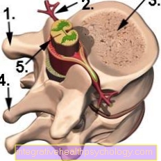
In addition to injuring blood vessels, organs can also be damaged, resulting in a hemothorax. This can affect all organs that are located within the chest. Especially injuries to the Mediastinum, a demarcated area in the center of the chest cavity, can create a hemothorax. The mediastinum contains the Thymus ("Bries") that windpipe, the esophagus and some Lymph nodes. The thymus recedes at a young age, but it can be injured in children, for example, when a bicycle falls or handlebar injuries.
Hemothorax after medical measures

In addition to the traumatic causes, a hemothorax can also be triggered by a medical measure. This includes the installation of a central venous catheter (CVC) in the jugular vein or a drainage as well as the puncture of the pleura, which is carried out for diagnostic or therapeutic reasons.
Since the attending physician proceeds according to a standardized scheme, it is possible that in the individual case the blood vessels of a patient run differently than normal and the blood vessels are punctured. The attending physician can also set the drainage or puncture needle incorrectly, resulting in a hemothorax.
In the "Thoracotomy"is a surgical procedure in which the chest cavity is opened. It can be operated on the lungs, on the heart or on the organs that are located within the mediastinum. Since a surgical procedure without bleeding is not possible At the end of the operation, drains are placed in the chest and on the affected operating area to drain the excess blood. If these drainages are insufficient, blood can also build up in the chest and thus lead to a hemothorax.
Symptoms
Symptoms vary depending on the amount of fluid retention. If there is heavy bleeding into the pleural space, it comes to Shortness of breathbecause the lungs can no longer develop properly due to the spatial restriction caused by the accumulation of blood. As a result of the impaired breathing it comes to one Lack of oxygen. The consequence of the lack of oxygen is a blue coloration of the skin ("cyanosis"), Dizziness, fainting, and muscle weakness.
In addition to the lack of oxygen, there is a lack of blood in the body's circulation, especially in the case of heavy bleeding. Due to the loss of blood, the human body reacts with a counter-regulation. Of the Blood pressure drops by the decreased blood volume, whereas the Pulse increases significantly. In addition, there is a so-called centralization of the blood. This means that the body conducts more blood through the vessels near the heart and the distant limbs such as fingers and toes are less well supplied with blood. This sustains the heart's action.
The urine output is also downregulated by the body in order to save as much fluid as possible. However, if the blood loss is too great, the condition may change Shocks develop.
diagnosis
X-ray for a hemothorax

A large area of shadowing can be seen in the X-ray image if a hemothorax is present. Depending on the extent of the injury, this can occur either immediately after the trauma or in the course of the next few hours. In addition to the hemothorax, an accompanying pneumothorax (air accumulation in the chest) can also be seen in the X-ray.
In addition, the examiner should look out for injuries to the spine and ribs. A diagnosis can be made quickly and inexpensively with an X-ray image, but there is a disadvantage in the radiation exposure from the X-ray device and in the fact that an accumulation of fluid can only be detected from around 200 ml.
Read more on the topic: Chest x-ray (chest x-ray)
Ultrasound for a hemothorax
The ultrasound examination can also be particularly good small accumulations of fluid from 50ml can be recognized. This simple and inexpensive method is particularly suitable for monitoring the progress of a hemothorax, as it can be carried out with the patient lying down and simply in the patient's bed.
With the ultrasound examination, complications can be identified and treated quickly and easily. However, it is not possible through the ultrasound examination to obtain an accurate representation of air-conducting structures and from the lung to produce, since air-containing rooms are difficult to represent. Accompanying injuries to these organs can possibly be overlooked.
CT for a hemothorax
Also the CT examination is a diagnostic option if a hemothorax is suspected. Computed tomographic imaging is the most accurate and the most detailed investigation method. In addition to air and fluid accumulations in the chest cavity, injuries to neighboring organs can also be detected.
Due to the cross-sectional imaging, the bony Structures can be recognized well and one or more fractures of the ribs, the sternum or the spine can also be excluded. The CT examination enables quick and non-invasive diagnostics, but the device has the disadvantage of being high Radiation exposure (about 1000 times higher than with normal X-rays).
therapy
To ensure targeted therapy, the cause of the hemothorax should first be determined. If this involves injuries to vessels or organs, these should be treated first in order to avoid major blood loss and to keep the accumulation of blood in the chest as low as possible.
The next step should be a so-called Chest drain be placed. This is a tube system that is placed from the outside between the two pleural leaves and directly into the effusion. This drainage should allow blood to drain out of the chest. In addition, a valve can be attached to the drainage, which pulls out the remaining liquid with a suction.
In addition to the dissipative function, a Flushing of the pleural space to remove any remnants of the effusion. Antibiotics can be added to the irrigation solution to minimize the risk of localized inflammation.
The thoracic drainage is placed through a 2-3 cm skin incision in a space between the ribs, the subcutaneous fatty tissue is displaced with blunt scissors. As soon as the pleura is opened, the drainage can be pushed into the chest cavity. It is fixed in place by a skin suture. The drainage is often located in the space between the 4th and 5th ribs on the side of the chest, this technique is used Bülau drainage called. However, it can also be created further above.
Please also read our article on this Chest drain.
If the hemothorax has arisen as a result of vascular or organ injuries or there is a traumatic injury to the chest from the outside, a Thoracotomy take place as therapy. This is a surgical opening of the chest. For this purpose, the patient is usually positioned on their side and the injury within the chest cavity can be surgically treated via various access routes. The hemothorax, i.e. the accumulation of blood, can also be completely suctioned off and cleared out via this access route. This avoids hardening of the effusion and the resulting sticking of the pleura sheets to one another.
During this operation it is also possible to put on a chest drain from the inside. In addition to these invasive measures, prophylactic antibiotic therapy should be carried out over a period of several weeks to prevent bacteria from settling in the chest cavity and the resulting inflammation.
Complications of a hemothorax
In the case of very heavy bleeding into the chest due to vascular or organ injuries, uncontrollable blood loss can occur and there is an imminent danger to life. Therefore, a hemothorax should be treated as quickly as possible by specialist staff or, as an initial measure, by an emergency doctor.
If survival is ensured, however, late complications of the hemothorax can still occur. A therapeutic or traumatic opening of the rib cage poses a risk of infection. These can take many different forms.
Pleurisy, i.e. inflammation of the pleura, can occur. This can result in numerous complications that may have long-term effects on breathing.
Above all, this includes the pleural rind. This is a gluing of the two sheets of the pleura, which is accompanied by a thickening and hardening. This happens especially after inflammation of the pleura, which is why the therapeutic administration of antibiotics is particularly important in the context of inflammation prophylaxis. The tissue of the pleural rind tends to contract over time, causing a loss of elasticity.
The lungs can no longer fully develop and the breathing volume decreases as a result. Therefore, a pleural skin should always be treated surgically or avoided in advance by clearing out the hemothorax.