Brain stem
Synonyms in the broadest sense
Truncus encephali
introduction
The brain stem too Truncus encepahli consists of the following components:
- Midbrain = mesencephalon
- Afterbrain = metencephalon from bridge (pons) and cerebellum (cerebellum)
- Elongated medulla = medulla oblangata
General
Of the Brain stem of Brain embraces this from top to bottom Midbrain, the bridge with IV. Cerebral ventricle as well as bordering on it Cerebellum and at the bottom that extended markwhich in the Spinal cord transforms. The brain stem also contains the cranial nerve nuclei of the third to twelfth cranial nerves.
Midbrain
The midbrain in the brain is 1.5 to 2 cm in size and divides from front to back into the two cerebral legs (Crura cerebri), the hood (Tegmentum) and the four-hill slab (Tectum). The IV cranial nerve (Trochlear nerve) out. Inside the aqueductus mesencephali (aquaeductus = water pipe) as a connection from III. and IV. ventricle. As a connection with the cerebellum, the upper cerebellar legs leave (Pedunculi cerebellares superiores) the midbrain.
Regarding the important nuclei of the midbrain, the central cave gray (Substantia grisea centralis = central gray matter), the reticular format ("Web-like formation", nerve cell network), the substantia nigra (black substance) with melanin-containing nerve cells and the iron-containing nucleus ruber (red core) to call.
Furthermore one finds the nuclei of the III. and IV. cranial nerves.
Also read our main article: The Midbrain
bridge
The Bridge of the brain has a structure comparable to the midbrain (brain stem): Bridge foot (front), Bridge hood (center) and Velum medullare (behind; velum = sail, medullare = medullary).
The bridge also has a connection to the cerebellum with the middle cerebellar legs (Pedunculi cerebllares medii).
The bridge hood (brain stem) also contains the Formatio reticularis, the Locus caeruleus as well as the cranial nerve nuclei of the cranial nerves V to VIII.
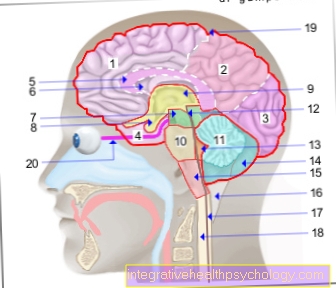
Illustration brain

Cerebrum (1st - 6th) = endbrain -
Telencephalon (Cerembrum)
- Frontal lobe - Frontal lobe
- Parietal lobe - Parietal lobe
- Occipital lobe -
Occipital lobe - Temporal lobe -
Temporal lobe - Bar - Corpus callosum
- Lateral ventricle -
Lateral ventricle - Midbrain - Mesencephalon
Diencephalon (8th and 9th) -
Diencephalon - Pituitary gland - Hypophysis
- Third ventricle -
Ventriculus tertius - Bridge - Pons
- Cerebellum - Cerebellum
- Midbrain aquifer -
Aqueductus mesencephali - Fourth ventricle - Ventriculus quartus
- Cerebellar hemisphere - Hemispherium cerebelli
- Elongated Mark -
Myelencephalon (Medulla oblongata) - Big cistern -
Cisterna cerebellomedullaris posterior - Central canal (of the spinal cord) -
Central canal - Spinal cord - Medulla spinalis
- External cerebral water space -
Subarachnoid space
(leptomeningeum) - Optic nerve - Optic nerve
Forebrain (Prosencephalon)
= Cerebrum + diencephalon
(1.-6. + 8.-9.)
Hindbrain (Metencephalon)
= Bridge + cerebellum (10th + 11th)
Hindbrain (Rhombencephalon)
= Bridge + cerebellum + elongated medulla
(10. + 11. + 15)
Brain stem (Truncus encephali)
= Midbrain + bridge + elongated medulla
(7. + 10. + 15.)
You can find an overview of all Dr-Gumpert images at: medical illustrations
Extended mark
The onion-shaped elongated pith (brain stem) is also built up in three layers, with an anterior and posterior area and a hood in between (Tegmentum). At the front of the extended marrow, the two pyramids with the pyramidal tracks run, the crossing of which (pyramid crossing) marks the end of the extended marrow. To the side of it are the olives, from which the lower cerebellar legs (pedunculi cerebellares inferiores) extend to the cerebellum. At the back of this part of the brain (brain stem) one finds the diamond pit; in addition to the reticular formation, the area postrema (Vomiting center) and various cranial nerve nuclei (of the VIII., IX., X. and XII. cranial nerves).
For more information, see: Extended mark
Cerebellum
The cerebellum as part of the brain rests on the brain stem at its rear and is connected to it via three cerebellar stalks (Pedunculi = little feet) connected. From the rest of the brain (cerebrum), under which the cerebellum is located, it is covered by a meningeal plate (Tentorium cerebelli, tentorium = tent) Cut.
The cerebellum is divided into the centrally located cerebellar worm (Vermis), which is surrounded on each side by a cerebellar hemisphere. Associated with the worm, the flocculus also belongs to it (flocculus = small tarpaulin). The surface of the cerebellum consists of furrows and convolutions that allow it to be divided into lobes.
For more information, see: Cerebellum
function

Overall that is Brain stem of the brain responsible for the regulation of vital processes such as sleep, breathing, Blood pressure level or micturition (Urination).
a) Midbrain: In the midbrain (brain stem) ascending pathways run to the brain and descending to the spinal cord. Furthermore, it takes with the central cave gray as part of the limbic system a task in the perception of pain. The Substantia nigra plays a role in motor system, of the Nucleus ruber in the coordination of muscle movements and muscle tension (muscle tone). On Visual processes (Eye movements, visual reflexes) as well as Auditory processes (auditory reflexes) the brain is involved via the four-hill plate.
b) bridge: Signals to the cerebellum are switched in the bridge.
c) extended mark: The elongated marrow (brain stem) forms with the Formatio reticularis an important reflex and coordination center of the brain. The Pyramid tracks (Corticospinal tract) establish the connection between the cerebral cortex and the spinal cord for the regulation of muscular work.
The Olive system forms as a switching station between the motor system and the cerebellum.
The Formatio reticularis (Brain stem) is found in the midbrain as well as in the bridge and in the elongated marrow. Their job is regulation vegetative and more affective Operations. Connections exist to limbic system (Mood), to Cerebral cortex (Awareness, falling asleep, waking up, arousal), to the Spinal cord (Pain suppression, motor processes), to sensory systems and to motor nerve stations (Muscle tension, coordination of stereotypical movements, control of eye movements). Furthermore, the reticular formation is involved in reflexes, whereby the Swallowing, sucking, corneal, vestibular reflex, oculomotor reflexes such as Secretory reflexes during digestion added.
d) cerebellum: The cerebellum, as part of the brain, plays a role in the coordination and regulation of muscular movements, including muscle tension (muscle tone) as well as the chronological sequence of movements. Together with the Labyrinth organ (Balance organ) it ensures balance. The cerebellum receives all of these tasks visual (regarding vision), auditory (regarding hearing), vestibular (the balance concerning), proprioceptive (concerning the depth sensitivity) and exteroceptive (Concerning touch, vibration, pressure, pain, temperature) information.


.jpg)


.jpg)