Hyaline cartilage
Synonyms in a broader sense
- Elastic cartilage
- Hyaline cartilage
English: Cartilage
definition
cartilage is a special form of connective tissue. A distinction is made between different forms of cartilage, which are adapted to the respective function.
Forms of cartilage are:
- hyaline cartilage
- elastic cartilage
- Fiber cartilage
Development of hyaline cartilage
Hyaline cartilage develops from the mesenchyme (form of connective tissue). At 45%, the proportion of collagen fibers is lower than that of fiber and elastic cartilage.
The collagen fibrils are masked by the glycosaminoglycans present in the basic substance. They are not visible in the light microscope image, since their refraction does not differ from that of the surroundings due to the low fiber density.
With the exception of articular cartilage, hyaline cartilage is one of them Cartilage skin (Perichondrium) covered. The innermost cell layer of the cartilage (stratumcellulare) the ability to form cartilage cells after growth is complete.
The Outer layer (Stratumfibrosum) consists predominantly of collagen fibers that absorb tensile forces that arise when the cartilage body is bent.
In this way, the cartilage retains a certain ability to regenerate even in adulthood. Nevertheless, the regenerative capacity of the hyaline articular cartilage is in principle low.
New cartilage can only be based on Perichondrium are formed. If the cartilage is missing, functional cartilage can no longer be built up after destruction as a result of inflammatory and degenerative joint diseases.
Chondrocytes (Cartilage cells) recede compared to the cartilage substance (extracellular matrix) in the vascular and nerve-free differentiated hyaline cartilage tissue, so that their volume percentage of cartilage cells is only between 1 and 10%.
Appointment with ?

I would be happy to advise you!
Who am I?
My name is I am a specialist in orthopedics and the founder of .
Various television programs and print media report regularly about my work. On HR television you can see me every 6 weeks live on "Hallo Hessen".
But now enough is indicated ;-)
In order to be able to treat successfully in orthopedics, a thorough examination, diagnosis and a medical history are required.
In our very economic world in particular, there is too little time to thoroughly grasp the complex diseases of orthopedics and thus initiate targeted treatment.
I don't want to join the ranks of "quick knife pullers".
The aim of any treatment is treatment without surgery.
Which therapy achieves the best results in the long term can only be determined after looking at all of the information (Examination, X-ray, ultrasound, MRI, etc.) be assessed.
You will find me:
- - orthopedic surgeons
14
You can make an appointment here.
Unfortunately, it is currently only possible to make an appointment with private health insurers. I hope for your understanding!
For more information about myself, see - Orthopedists.
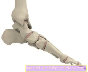
Illustration of cartilage damage

- Articular cartilage
(hyaline cartilage) -
Cartilago articularis - Cartilage remodeling zone
in bones -
Zona ossificationis - Joint body (articular gnar
of the femur) -
Femoral condyle - Femur -
Femur - Articular cartilage -
Cartilago articularis - Outer band -
Ligamentum collaterale fibulare - Outer meniscus -
Lateral meniscus - Inner meniscus -
Meniscus medialis - Fibula - Fibula
- Shin - Tibia
You can find an overview of all Dr-Gumpert images at: medical illustrations
Structure of hyaline cartilage
Hyaline cartilage when fresh looks bluish milky and appears transparent in thin slices. The cartilage substance (extracellular matrix) of the hylinen cartilage has a high water content of approx. 70%.
The dry substance of the cartilage consists of (structure):
- Proteoglycans
- Collagens
- Glycoproteins
- Lipids
and - Electrolytes.
Proteoglycans and Type II collagen fibers do with each 45% the main mass.
As the main proteoglycan of hyaline cartilage, the aggrecan forms together with the Hyaluronic acid the actual basic substance of the cartilage tissue.
Due to the high negative charge density of the glycosaminoglycan side chains, the aggrecan has a high reversible water binding capacity. This is explained by the partially positive charge of the water molecule as a dipole.
As a result, the water-laden glycosaminoglycans repel each other and build up a tissue-specific internal pressure (Swelling pressure of the cartilage) on who is about the tensile tensioning of the collagen fibers is held.
In free aqueous solution the Rejection of proteoaminoglycans expand huge. The proteoaminoglycans are retained by the collagen fibers of the extracellular matrix. The proteoaminoglycans can with Springs which are curbed and compressed by the collagen fibrils.
The high compressive elasticity comes about because the proteoaminoglycans allow a further compression, but immediately after the compression they expand again as much as the collagen fibrils allow.
In parallel, water is added compression displaced and tightened again on decompression. This Flexing of the articular cartilage is important for Nutrition of the cartilage.
The function of the cartilage depends on the one hand on the quantitative and qualitative composition of the proteoglycans and their GAG chains and on the other hand on the orderly structure of the collagen fibrils and their structure.
Both can become ineffective with increasing age, which is particularly noticeable in the joint cartilage in the form of symptoms in the joint.
Function of hyaline cartilage
In normal Joints the ends of the bones are covered with hyaline cartilage. In the articular cartilage, the collagen fibrils run like an arc. They pull up radially from the deepest zone, then bend in the tangential course and pull down again.
This creates one from top to bottom Zoning. In the Tangential zone the fibrils run tangential to the surface and are oriented in the direction of the greatest tensile stress.
Then one follows Transition zone and a Radial zone. In the zone of mineralized cartilage the fibrils are anchored and the zone is with the one beginning below bone interlocked. The arrangement is closely linked to the respective function.