Atlas
introduction
The atlas is the first cervical vertebra and the next to the skull of the spine. Because of this, he bears the burden of the entire skull.
It is also called "Nod“Because it enables nodding due to its structure and the attached muscles.
anatomy
Due to its special position and its special function, the atlas, like the second cervical vertebra (Axis) structured differently than all other vertebral bodies. The atlas forms a functional unit with the axis and consists of a small (ventral) and a large rear (dorsal) vertebral arch.
These vertebral arches each have a small bony attachment, the smaller one Anterior tubercle and the bigger one Posterior tubercle.
On the inside of the anterior vertebral arch is a small pit that Fovea dentis. This serves as a joint connection to the second cervical vertebra, the Dens axis.
On each side there is a thickened bony structure that Massea laterales.
These show a concave articular surface towards the top (Facies articularis superior), which serves as an articulation to the occiput.
Two further articular surfaces lie down on the massea laterales, the Inferior articular facies. These are used to connect to the Axis.
In the middle there is a large hole that Vertebral foramen. This is used to pass through the spinal cord.
On each side there is a smaller protrusion of bone, the Transverse process. In this there is a small hole that Foramen transversarium. It leads the Vertebral arterygoing through the occipital hole (Foramen magnum) enters the head.
The various bony protrusions serve as the origin and starting point for the prevertebral muscles and thereby enable the head to move.
Illustration of the cervical spine

Cervical spine (red)
- First cervical vertebra (carrier) -
Atlas - Second cervical vertebra (turner) -
Axis - Seventh cervical vertebra -
Vertebra prominent - First thoracic vertebra -
Vertebra thoracica I - Twelfth thoracic vertebra -
Vertebra thoracica XII - First lumbar vertebra -
Vertebra lumbalis I - Fifth lumbar vertebra -
Vertebra lumbalis V - Lumbar cruciate ligament kink -
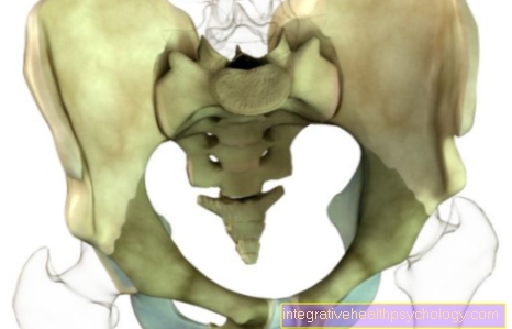
Promontory - Sacrum - Sacrum
- Tailbone - Os coccygis
You can find an overview of all Dr-Gumpert images at: medical illustrations
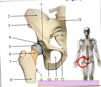
Joints
The atlas is the central element of the two head joints.
On the one hand, it forms the atlanto-occipital joint, which is the connection between the skull and the cervical spine. This joint enables flexion (Flexion), Elongation (Extension) and sideways movement of the head.
The atlantoaxial joint is the articulated connection between the first and second cervical vertebrae. It enables the head to be rotated.
Appointment with a back specialist?

I would be happy to advise you!
Who am I?
My name is I am a specialist in orthopedics and the founder of .
Various television programs and print media report regularly about my work. On HR television you can see me live every 6 weeks on "Hallo Hessen".
But now enough is indicated ;-)
The spine is difficult to treat. On the one hand it is exposed to high mechanical loads, on the other hand it has great mobility.
The treatment of the spine (e.g. herniated disc, facet syndrome, foramen stenosis, etc.) therefore requires a lot of experience.
I focus on a wide variety of diseases of the spine.
The aim of all treatment is treatment without surgery.
Which therapy achieves the best results in the long term can only be determined after looking at all of the information (Examination, X-ray, ultrasound, MRI, etc.) be assessed.
You can find me in:
- - your orthopedic surgeon
14
Directly to the online appointment arrangement
Unfortunately, it is currently only possible to make an appointment with private health insurers. I hope for your understanding!
You can find more information about me at


-operation.jpg)