Tibialis posterior tendon
definition
Tendons are stable, partially stretchable connections between muscles and bones.
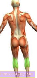
The tibialis posterior tendon connects the posterior tibialis muscle in the lower leg with the bone attachments under the foot. A movement of the muscle is passed on to the foot via the tendon and leads to a flexion of the sole of the foot, a pulling up of the inside of the foot and a pulling up of the foot in general.
Tendons can become irritated or tear under heavy strain. The tibialis posterior reflex can also be tested on the tendon.

Function of the tibialis posterior tendon
Tendons are connecting structures between muscles and bones. They pass on the strength of the muscles to the bones and thus cause the joints to move. The tendon of the posterior tibialis muscle transfers force to the bones in the foot. This causes the foot to tilt inwards (supination) and flexion of the soles of the feet and toes. These movements are called supination and plantar flexion.
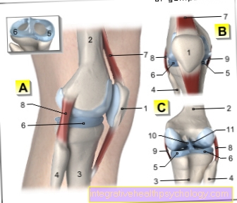
Tendons also stabilize the large joints, in this case the ankle, and can also give shape to other structures. The tendon of the posterior tibialis muscle helps stabilize the transverse arch of the foot and thus prevents splayfoot. Tendons can also store strength and release it later. With regular exercise, such as running, the tibialis posterior tendon can store strength and relieve the calf muscles, resulting in a smoother movement. This can also protect the joints and bones.
Tendons are under heavy strain for a lifetime and can therefore show functional losses in old age. This manifests itself in pain and a greater risk of injury. Cracks, inflammation or malformations can also prevent the tendon from performing its function or not being able to perform its function sufficiently.
Course of the tibialis posterior tendon
The tibialis posterior tendon begins on the lower leg at the end of the posterior tibialis muscle and crosses under the tendon of the long flexor toe muscle. This is called the Chiasma Crurale or the crossing of the lower legs. Then the tendon runs through the tarsal tunnel, a connective tissue supporting apparatus of the ankle. At the ankle, the tendon runs along the inner ankle and can be used just below or above the ankle to test the tibialis posterior reflex. The tendon splits into several end pieces and attaches to the sphenoid bone and the navicular bone. These are tarsal bones. Further approaches are the individual toes, which can be bent by the posterior tibialis muscle.
If you are also interested in the anatomy of the posterior tibialis muscle, please also read: Tibialis posterior muscle
Diseases of the Tibialis Posterior Tendon
The tendon of the tibialis posterior muscle can become inflamed if it is severely irritated or rupture or tear under sudden, heavy loads.
Tendon pain
In most cases, tendon pain occurs during tendon stress. However, pain is only a symptom of another damage and not the disease itself. The pain can be the result of traumatic damage, i.e. a rupture or stretching when twisting an ankle, or it can be triggered by inflammation of the tendon or the tendon sheath. Those affected can try to contain the pain first by taking a rest and taking painkillers. If the pain persists, a doctor should be consulted.
Inflammation of the tendon
The tendon of the posterior tibialis muscle runs over several joints and has to withstand extreme loads. The tendon is irritated with every single step. This irritation can lead to an inflammation of the tendon, especially with increased stress, such as in competitive athletes. The tendon sheaths can also become inflamed and sticky if they are subjected to excessive stress. In both cases, those affected have severe pain, which is aggravated by the strain on the tendon, for example when running. The incidence of tendinitis increases in old age, as the tendons can be affected by a degenerative change. Inflammation of the tendons or tendon sheaths can spread to surrounding structures.
In most cases, conservative therapy is sufficient. The tendon is immobilized and brought back to normal function with certain physiotherapeutic exercises. During this time, the affected tendon should be spared. Painkillers and anti-inflammatory drugs are also used. In the event of persistent severe pain, surgical removal of the affected tendon piece can be considered. The tendon is removed piece by piece and the stump is sewn back together.
For more information, see: Tendonitis of the tibialis posterior tendon
Rupture and tear of the tendon
A tendon is exposed to strong mechanical stress. Usually tendons are built to do just that and can withstand great forces, but sudden, heavy stress can cause the tendon to tear. With age, the elasticity of the tendons also decreases and the likelihood of rupture increases. A torn tendon is very painful for the person concerned and leads to a complete loss of function of the affected muscle. In some cases, conservative treatment is sufficient, while a complete transection often requires surgery to sew the tendon back together.
For more information on the torn tibialis tendon, as well as other tendon tears, see: Tendon tear
Tibialis Posterior Syndrome
Tibialis posterior syndrome is a progressive disease of the tendon of the posterior tibialis muscle. This is always caused by a degenerative (degenerative) change in the tendon and occurs more frequently in old age. Women are three times more likely to be affected than men. The increasing degeneration gradually leads to functional restrictions. Those affected find it increasingly difficult to move their foot inward and the sole of the foot to bend less and less. Since the tendon of the posterior tibialis muscle also stabilizes the arch of the foot, a flat foot that is inclined outwards and the associated damage to the joints occurs later on, as the running movement can no longer be sufficiently cushioned.
The first approach to therapy is usually conservative. Those affected should take care of their feet and receive physiotherapy. Shoe insoles are also recommended as a support. Pain relievers and anti-inflammatory drugs can also be used. Surgical therapy is an option for more severe courses. The inflamed or dead tissue is removed and the tendon is reconstructed with healthy tendons. For some people, the bone also needs to be treated.
The tarsal tunnel syndrome is caused by a nerve constriction in the course behind the inner ankle (tarsal tunnel). It also causes pain in the foot. If you want to learn more about it, please also read: Tarsal tunnel syndrome
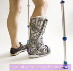
How do you wallpaper the tibialis posterior tendon?
Since the tendon of the posterior tibialis muscle passes several joints, all directions of movement of the tendon must be taped. The first pulling direction runs straight down the inside of the lower leg to the sole of the foot. The second pulling direction starts at the front of the lower leg and pulls across to the same point on the sole of the foot as the first tape. The third tape runs around the heel to the front third of the foot. In this way, all parts of the tendon are stabilized.
For more information, we recommend our website: Taping the ankle
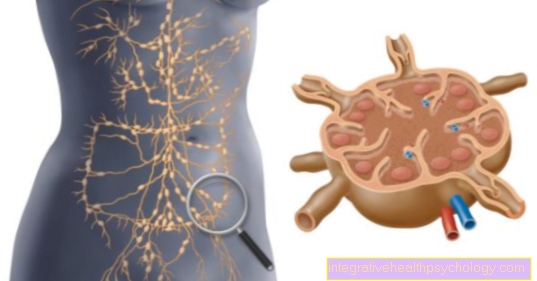
Microscopic structure of a tendon
The structure of body parts is always divided into a microscopic part and a part that is visible to the eye. Microscopically, a tendon is a fibrous connective tissue that is only supplied by a few blood vessels. The end of the tendon is anchored directly to the tendon fibers in the bone or in the periosteum. The tendon itself is surrounded by a thin skin. Since the tendon of the posterior tibialis muscle runs over several joints, it is additionally protected by tendon sheaths.