Cerebellar atrophy
introduction
The brain is made up of different parts, including the cerebellum. It plays an important role in coordinating and fine-tuning the movements of different muscles and balance.
It is also believed to be involved in many cognitive and emotional skills. It is found in the posterior fossa. It lies under the cerebrum and behind the brain stem.
The cerebellum is covered by the cerebellar tent which separates the cerebellum from the cerebrum.

Classification of cerebellar atrophy
Cerebellar atrophy is a demonstrable loss of tissue in the cerebellum. This means that the cerebellum becomes smaller and can no longer fully fulfill its tasks.
It is divided into three areas, each with different functions. Depending on the extent to which one or more areas are affected, the functional failures are different.
Depending on the causes, certain cerebellar areas tend to shrink. Depending on the cause, an increased shrinkage of the cerebellar cortex or the cerebellar medulla is observed.
The precise implications for the mass of cerebellar tissue shrinkage are controversial.
causes
The causes of tissue atrophy in the cerebellum are very diverse and can occur at any age. The causes and the resulting diagnosis and therapy can be classified into three groups of affected persons:
- Affected people who become ill before the age of 25 and whose siblings are also affected or if the cerebellar atrophy occurs sporadically.
- Affected people in whom the symptoms of cerebellar atrophy vary and one parent is affected.
- Affected people who fall ill after their 40th year of life and if the disease occurs sporadically.
Furthermore, the cerebellar atrophy can be divided into three forms, in which the affected groups are represented differently:
- Inherited forms: Usually this is an autosomal dominant inheritance. A further distinction is made here depending on the accompanying symptoms and depending on which cerebellar area or, if applicable, which other brain areas are most affected.
- Symptomatic forms are usually caused by a tumor disease, by prion viruses or by toxic substances such as alcohol or medication (e.g. cytostatics).
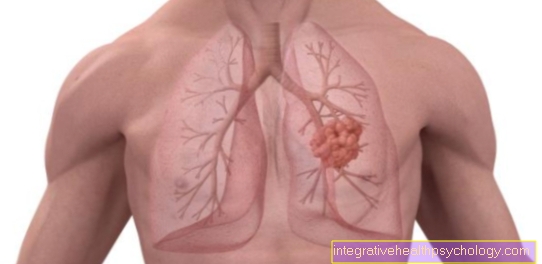
It is assumed that antibodies are initially directed against the tumor tissue. Often the cerebellar atrophy can be seen before the tumor disease. Often they are small-cell lung carcinomas or gynecological tumors. - Sporadic forms are often found in the context of multiple tissue atrophy. Otherwise, one speaks of sporadic forms if the other two groups of causes mentioned can be excluded.
Read more on the subject at:
- Cerebellar damage
- Brain atrophy
alcohol
Cerebellar atrophy can be caused by alcohol (symptomatic form). Some researchers suggest that the cerebellum in particular is sensitive to the toxic effects of alcohol.
In the context of chronic alcoholism, the tissue atrophy in the cerebellum can manifest itself in the form of the Charcot triad characteristic of cerebellar disorders: double vision, balance or coordination disorders, and a speech disorder. Eye tremors are observed less often.
Depending on the extent and the affected cerebellar area, the symptoms show up differently. Some researchers observed tissue shrinkage in the cerebellum caused by alcohol, in particular a reduction in size of the cerebellar worm, which is responsible for Vestibulocerebellum heard. The vestibulocerebellum is a part of the cerebellum that receives information from the balance organs and is responsible for coordinating head posture, head and eye movements.
As a result, there are often limitations and dysfunction in the corresponding cerebellar areas.
It is also suspected that the gray matter in alcohol-related cerebellar atrophy is particularly affected. For example, speech disorders in cerebellar atrophy, which is caused by alcohol, express themselves differently than speech disorders in cerebellar damage caused by other diseases, such as Multiple sclerosis.
In the former, the speech disorder is characterized by unclear pronunciation and changing volume. With the latter one often observes a slow, choppy, slurred speech melody.
Both the toxic effects of the alcohol itself and the presumably resulting thiamine and vitamin B deficiency can cause cerebellar atrophy.
A general laboratory test and a determination of the vitamins, as well as a determination of the alcohol abuse markers, e.g. the carbohydrate-deficient transferrin (CTD) can provide information. Further cerebellar atrophy can be stopped by abstaining from alcohol and giving the missing vitamins. Treatment for alcohol sickness is recommended. If alcohol is continued to be consumed, the cerebellar atrophy progresses.
The effect of alcohol during pregnancy on the unborn child with regard to the growth of the cerebellum and the growth and functionality of other structures must also be viewed very critically. Alcohol during pregnancy can cause cerebellar atrophy, among other things. Therefore, alcohol consumption during pregnancy is strongly warned.
Read more on the subject at: Consequences of alcohol
Symptoms
Depending on the affected cerebellar area and the extent of the tissue atrophy, characteristic symptoms of cerebellar atrophy occur. The cerebellum can be divided into three sections with different functions. The Vestibulocerebellum mainly processes information from the equilibrium organs and is responsible for coordinating head and eye movements. The Spinocerebellum regulates walking and standing and the pontocerebellum is used for fine regulation of motor skills and the correct execution of movements.
If the vestibulocerebellum is affected, the person concerned usually suffers from dizziness, balance disorders, unsteadiness, speech disorders and eye movement disorders, sometimes with double vision and eye tremors.
The speech disorder is characterized by unclear pronunciation and changing volume. This cerebellar area is often affected when alcohol is the cause of cerebellar atrophy.
If the spinocerebellum is affected, there is usually an unsteady stance and gait, known as so-called stance and gait ataxias.
When the pontocerebellum shrinks, people experience limited, aimless grasping and tremors during these movements. There is also a delayed braking of the muscle opponents, as well as coordination disorders, speech disorders and an inability to perform rapid sequences of movements.
The speech disorder is characterized by a slow, slurred, choppy speech melody.
This area of the cerebellum is often affected when the damage to the cerebellum is caused by a disease such as multiple sclerosis.
In the case of cerebellar atrophy, all of the mentioned cerebellar areas can also be affected together. In relation to cerebellar atrophy, researchers also suspect connections between classical conditioning, phobias, and cognitive and emotional abilities with effects at the cerebellar level. As a result, cerebellar atrophy can also limit and affect these capabilities and areas.
Read more on the subject at: Functions of the cerebellum
diagnosis
A survey of the person concerned and a clinical examination can provide evidence of cerebellar atrophy. The doctor asks about the patient's medical history and biography and checks them Movement, coordination, speaking, and eye movements.
With imaging diagnostics the extent of the cerebellar atrophy can be measured more precisely. If there is a genetic cause, genetic diagnostics should be carried out and comorbidities should be inquired about.
A blood test is necessary for alcohol-related cerebellar atrophy.
MRI of the brain for cerebellar atrophy
With the help of an MRI examination, also known as magnetic resonance tomography, the exact extent of the small reduction can be visualized. Surrounding structures can also be examined for participation.
The magnetic resonance examination enables a kind of "dynamic view" of the cerebellum and other brain regions. The functional MRI can provide insights into events over time and the activity of the cerebellar areas. Depending on the objective of the examination, contrast media are also used.
The MRI is X-ray free and there are usually no medical risks if the regulations presented during the examination are followed. For example, no clothing or jewelry containing metal may be worn.
Risks, procedure of the examination, functional principle, procedures, indications and duration of the MRI examination of the brain correspond by and large to the explanations of the general MRI examination.
After an MRI of the brain, the cross-sectional images of the brain can then be displayed on each level on the PC monitor.This allows structures of the cerebellum to be made relatively detailed and cerebellar atrophies to be made visible relatively precisely.
You can also read the relevant information at: MRI of the brain
Illustration brain

Cerebrum (1st - 6th) = endbrain -
Telencephalon (Cerembrum)
- Frontal lobe - Frontal lobe
- Parietal lobe - Parietal lobe
- Occipital lobe -
Occipital lobe - Temporal lobe -
Temporal lobe - Bar - Corpus callosum
- Lateral ventricle -
Lateral ventricle - Midbrain - Mesencephalon
Diencephalon (8th and 9th) -
Diencephalon - Pituitary gland - Hypophysis
- Third ventricle -
Ventriculus tertius - Bridge - Pons
- Cerebellum - Cerebellum
- Midbrain aquifer -
Aqueductus mesencephali - Fourth ventricle - Ventriculus quartus
- Cerebellar hemisphere - Hemispherium cerebelli
- Elongated Mark -
Myelencephalon (Medulla oblongata) - Big cistern -
Cisterna cerebellomedullaris posterior - Central canal (of the spinal cord) -
Central canal - Spinal cord - Medulla spinalis
- External cerebral water space -
Subarachnoid space
(leptomeningeum) - Optic nerve - Optic nerve
Forebrain (Prosencephalon)
= Cerebrum + diencephalon
(1.-6. + 8.-9.)
Hindbrain (Metencephalon)
= Bridge + cerebellum (10th + 11th)
Hindbrain (Rhombencephalon)
= Bridge + cerebellum + elongated medulla
(10. + 11. + 15)
Brain stem (Truncus encephali)
= Midbrain + bridge + elongated medulla
(7. + 10. + 15.)
You can find an overview of all Dr-Gumpert images at: medical illustrations
therapy
If there is an underlying disease (in the symptomatic form), it should be treated first.
Depending on the cause, (additionally) specific, individually tailored measures are recommended.
Scientific studies on the effectiveness of drug treatment for the various complaints are still ongoing. One study showed success in treating the Ataxias (e.g. gait uncertainties) observed with Riluzol®. However, since the number of participants in the study was small, it should be viewed critically and expanded through further studies.
For example, propanolol, carbamazepine, topiramate and clonazepam have been used to treat tremors during targeted movements. In some cases, however, a worsening of the symptoms could be observed.
In the case of eye movement disorders, a distinction is made between treating eye tremors and treating double vision.
If the eyes are trembling, guidelines recommend treatment with baclofen, gabapentin and 3,4-diaminopyridine. In the case of double vision, prismatic glasses can sometimes help.
Targeted eye muscle training also helps some sufferers.
According to the guideline, various medications are recommended to alleviate the symptoms, depending on other accompanying symptoms. The side effects must be taken into account and an individually tailored treatment with the help of a specialist should be sought.
Targeted occupational, physiotherapy and speech therapy are recommended to maintain independence and participation in everyday life. The contents of the therapies should be based on the complaints, resources and goals of the person affected and should also include advice for relatives and aids.
What is the life expectancy?
Since the reason for cerebellar atrophy is not always the same, there is nothing general to say about life expectancy. You have to look at each cause individually. In general, one can say that the life expectancy of the symptomatic cause is determined by the actual underlying disease. These include, for example, the paraneoplastic symptom in small cell lung cancer with a 5-year survival rate of approx. 15%, or in ovarian cancer (Ovarian cancer) with a 5-year survival rate of around 40%. Alcohol sickness also falls under the point. Studies have found that alcoholics live around 20 years shorter.
No statement on life expectancy can be made about the hereditary, i.e. hereditary causes, since no clear tendencies could be determined even in the subgroups of hereditary cerebellar atrophies. However, one can generally make statements about the quality of life. Cerebellar atrophy is usually a slow, chronic, deteriorating disease. The hereditary form can only be treated symptomatically. Many different specialist departments, such as speech therapy, occupational therapy and physiotherapy, help. It is hoped that this will delay the progression of the disease. Due to the unsteady gait, a wheelchair is often required after many years, but this varies from patient to patient. What is special about the symptomatic form of the disease is that abstinence from the toxin (e.g. alcohol) does not lead to any further breakdown of the brain tissue.
course
The course of cerebellar atrophy is individual and does not give a cure. Adequate lifestyle choices can delay the progression of the disease.
In the case of alcohol-related cerebellar atrophy, this includes, for example Alcohol abstinence, the addition of missing vitamins and treatment for alcoholism.
Active participation in targeted occupational therapy, physiotherapy and speech therapy can promote the course of cerebellar atrophy.
A harmonious environment (kindergarten, school, work, living area, leisure activities) can also delay the progression of cerebellar atrophy. On the other hand, passive interaction and an inappropriate lifestyle (e.g. continued consumption of alcohol) promote the atrophy of the cerebellum.
It is still largely unknown to what extent other brain areas can take on the tasks of a dwindling cerebellum. This is discussed very controversially by many researchers.
Consequences of cerebellar atrophy
As mentioned above, the disease has a significant impact on the quality of life of patients.
Patients affected by the hereditary form spend a lot of time on therapy without a chance of recovery. Genetic counseling should be given if these patients plan to have offspring. Such a chronic illness is often a burden on the psyche that should not be neglected.
As already explained, the cause of the symptomatic form is different. Alcoholics need to make a lifestyle to stop further deterioration. For cancer patients, cerebellar atrophy is a secondary diagnosis and they have to face the strenuous cancer therapy. But as I said, not every patient is equally severely affected. In the early stages in particular, it is often still possible to pursue a career and organize leisure time.
Duration
The Cerebellar atropy cannot be cured. It can only slow the progression of the disease. How long the illness lasts is very individual and depends on the underlying illness and individual living conditions.
Many individual factors play a role, which is why information on the duration and progression of cerebellar atrophy is very unreliable.
Cerebellar atrophy and dementia
There are studies on the autosomal dominant hereditary cerebellar atrophy (ADCA - autosomal dominant cerebellar ataxia) and a connection with dementia.
Only subtype 1 is said to have an association with mild dementia. Above all, attention and the ability to learn are disturbed. The subtypes of the hereditary form are determined through a genetic test.
Also read:
- Dementia test
- Drugs for dementia
Cerebellar atrophy in children
A Cerebellar atrophy in children can be idiopathic in origin, meaning that the cause of the disease is unclear. But it can also be genetic.
A few years ago it was discovered that certain drugs that were used against viral infections and that block DNA synthesis, cerebellar atrophy and in young children Cerebellar damage have caused.
The synthesis of DNA is at the Nerve cell formation in the cerebellum not yet completed in toddler age. Therefore, such drugs can potentially interfere with cerebellar development.
The same symptoms, diagnostic procedures and therapies apply to cerebellar atrophy in children as in adults. The occupational, physiotherapy and speech therapy should be carried out individually and in a child-friendly manner.
It will be a possible early start of therapy recommended, which should include extensive parenting advice.
At best, educators from integrative institutions or from special / support kindergartens or schools should also be informed and involved.

.jpg)
.jpg)