Bone density measurement using the DEXA method
The DXA measurement, also known as dual X-ray absorptiometry, is a diagnostic procedure that is mainly used to measure bone density. It can also determine the body composition and thus determine the percentage of body fat, lean mass and bone mass of the person examined. The technology behind the procedure is based on X-rays.
In recent years, the DXA measurement has been used very often, especially in the area of determining bone density. The measurement can identify existing and incipient osteoporosis and initiate treatments.

principle
The principle of DXA measurement is based on the Principle of the X-ray image. However, different from a normal X-ray multiple images made what differ in their radiation density. Using this method, the Density of the bone can be calculated exactly. The calculation is carried out by a computer, usually connected to the device.
On the one hand, the ideal place to do this is Hip bones and on the other hand the Lumbar spine. It can other body parts as well can be examined, the accuracy of the measurement results decreasing in this case.
Even if osteoporosis affects the whole body, the measurements will be preferably on the hip joint and spine made. The reason is in the comparability the implementation and calculation of the values for other tested persons and study results as well as the accuracy the Results.
You can find more information on our website Bone density measurement.
Indications
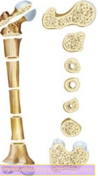
The most common indication for performing a DXA measurement is Suspected osteoporosis. The disease is statistically significant more often women than men, with women often following the menopause be diagnosed with the condition. Children are usually not affected. Osteoporosis goes with one Loss of calcium in the bones hand in hand. The structure and stability of the bone changes, the Bone becomes thinner and the danger of one Fracture becomes significantly larger.
Even with already diagnosed osteoporosis or other diseases that are associated with a changed bone density, the DXA measurement can be used for the process control be used.
A DXA measurement can provide information about how high the risk is spontaneous bone fracture to suffer. The risk arises from age, body weight, any previous bone fractures, family history and certain behaviors like that Smoke and the Consumption of alcohol and other drugs. Taking these factors into account as well as that Create a DXA file for the person concerned, enables the attending physician to draw up a risk profile.
The DXA measurement is recommended for:
- Women after her menopauseif no estrogen is taken, or risk factors are present.
- when a personal history of broken bones and possibly a family history is known.
- if clinical symptoms are known which can be associated with loss of bone density.
- if certain drugs which can adversely affect the density of the bone.
- when a Type I diabetes, one Liver- or Kidney disease or have a family history of osteoporosis.
- when a Overactive thyroid present (Hyperthyroidism).
- when a Overactive parathyroid gland present (Hyperparathyroidism).
- when a Broken bone took place, though just a slight trauma occurred in which no fracture would have been expected.
- if imaging shows a Fracture of the spineor other signs of osteoporosis have been detected.
Please also read our page Diagnosis of osteoporosis.
Frequency distribution
Osteoporosis is a disease that has been around for the last few years significantly increasing numbers could post. The World health organization WHO classifies the disease as one of the ten most important diseases of today. Studies go by about 6.3 million people in Germany who suffer from osteoporosis. The best method, which was also classified by the WHO as the gold standard, for the early detection of osteoporosis and which could show good results in follow-up controls, is the DXA measurement.
execution
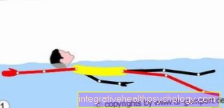
The DXA measurement is usually carried out by one Specialist in orthopedics or radiology, but can also be done in a hospital. The special devices allow the measurement while the patient is in motion in a horizontal position lies. The X-ray tube is located under the patient who detectorwhich detects the transmitted rays is located above the patient. So that the spine can be measured as precisely as possible, the Legs slightly elevated become.
It is important that the person examined is not movedso that the results of the measurement are accurate. The examination takes, depending on the device and body parts to be examined, about 10 to 30 minutes. The patient does not notice the examination. The DXA measurement remains in many cases no one-time examination, but is used several times for a follow-up check. The usual intervals between the examinations depend on the disease 6 months to 2 years.
advantages
The DXA measurement is one simple, fast and non-invasive measuring method. It won't anesthesia or Local anesthesia required to perform the measurement. The radiation density that is required is very small and only a fraction of the amount of radiation that is required, for example, with a Computed Tomography is blasted on the body.
The DXA method is that most accurate method availablewhich can reliably diagnose a disease of osteoporosis and is also suitable for the Risk of spontaneous bone fracture to investigate. In addition, devices with which a DXA measurement can be carried out are now available Very commonso that the implementation is very practical for the patient and for the doctor. Typically, x-rays have been in the doses used for diagnostics no side effects on the human body and can therefore be classified as harmless for most people.
disadvantage
Despite the low dose of radiation exposure which is necessary for the implementation of the DXA measurement remains some residual riskthat radiation can cause damage. In a healthy, adult person, the risk is low, and in most cases outweigh the benefits the method versus the low risk to the body. However, this risk results in Children and young people and especially pregnant women cannot be examined with the DXA measurement.
The attending physician should therefore be advised before carrying out the measurement of a possible pregnancy necessarily be taught.
Limits
The DXA measurement cannot determine exactly which patient is at what point possibly a fracture gets, it's just possible that relative risk to identify the data subject.
The accuracy and feasibility of the DXA measurement is with people who Bone changes in the spine area, or a previous spine surgery may not have given more. Likewise, existing fractures can affect the accuracy of the examination, which is why in these cases usually a computed tomography is indexed.
Alternatives
The DXA measurement represents that on commonly used procedures for the determination of the bone density. However, for certain reasons other methods are used for the measurement.
- An alternative to the DXA measurement is the so-called quantitative computed tomography (QCT). The advantage of this method is the possibility of a 3D illustration of the body to make. The accuracy despite one significantly higher radiation exposure However, this measurement is a not insignificant disadvantage.
- Another method that also shows the body in a 3D image is that peripheral quantitative computed tomography (pQCT). As with this method, however only on peripheral parts of the body Measurements are carried out, studies show that the measurement results do not come close to the accuracy of the results of a DXA measurement and thus changes that are associated with osteoporosis, for example, do not document adequately can.
- There is also another method which Completely without harmful X-rays gets by is the so-called quantitative ultrasound examination (QUS). However, this method is only partially suitable for the process control of diseases with altered Bone densitybecause changes cannot be assessed.
costs

The DXA measurement to determine bone density will be partially paid by health insurance. The statutory health insurance companies only pay for a bone density measurement if this is carried out using a DXA measurement. All other examination methods are generally used if the bone density is to be determined, not paid by the statutory health insurance.
The DXA measurement is accepted when a Broken bone, which one without previous trauma arose, which could explain the break. The examination is also paid for, if concrete findings on the medical side, which one osteoporosis suggest. In all other cases the investigation must self paid become. The cost is approx between 40 and 50 €.


.jpg)