Rectum
Synonyms
Rectum, rectum
definition

The rectum is part of the last section of the large intestine (colon). Together with the anal canal (Canalis analis), the rectum is used for stool elimination (bowel evacuation, defecation).
construction
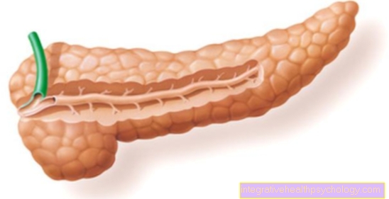
The Rectum is about 12-18 cm long, although this can vary from person to person. The name rectum is a bit misleading for the rectum, as the rectum is not straight but has curves in two planes.
In the side view, the rectum has two curves, the so-called Sacral flexure and the Perineal flexure. The sacral flexure points more in the direction of the sacrum (os sacrum), while the perineal flexure curves more in the direction of the abdominal wall, i.e. the anterior wall.
You can also see from the front Bends of the rectum, each of which dodges sideways. These bends are called Flexurae laterales designated. There are three lateral flexures. Opposite each bend in the mucous membrane of the rectum there is a corresponding one wrinkle (Plicae transversae recti). Of these three folds of the mucous membrane, the middle fold is particularly important. This middle fold of the mucous membrane is also called Kohlrausch fold designated.
The Kohlrausch fold is the most pronounced of the three folds and protrudes 6-7 cm into the intestinal lumen. The Kohlrausch fold marks that End of the rectal ampulla. The ampulla recti extends below the Kohlrausch fold and is the lowest section of the rectum. Clinically it is important that with one digital rectal exam (Palpation examination in which the doctor palpates the rectum with his finger) can be palpated approximately up to the Kohlrausch fold. This means that hardening such as tumors can be diagnosed manually here.
Below the ampulla recti limits the Anorectal junction the transition from rectum to Anal canal.
The anal canal is about 3 - 4 cm long and represents the very last part of the gastrointestinal tract. The end of the anal canal ends as anus between the two buttocks to the outside.
The wall structure of the rectum is three shifts. The outermost layer is dated peritoneum (Peritoneum) and fascia formed. The middle layer is the muscle layer. This consists of one Longitudinal muscles and one Circular muscles. The circular muscles are mainly in the area of the anal canal as the sphincter ani internus muscle (Internal sphincter) reinforced. The innermost layer in the wall structure is the mucous membrane. This lines the inside of the rectum.
Illustration of the rectum

- Rectum - Rectum
- Anal canal -
Canalis analis - Upper rectal artery
Superior rectal artery - Rectal vein plexus -
Plexus venosus rectalis - Rectum-anal canal boundary -
Anorectal junction - After lifter -
Levator ani muscle - Posterior columns -
Columnae anales - After pens -
Sinus anales - Internal anal sphincter -
M. sphincter ani internus - External anal sphincter -
External anal sphincter muscle - Anal canal-skin boundary -
Linea anocutanea - nach - anus
- Aft ridge line -
Linea pectinata - Sacrum - Sacrum
- Prostate -
prostate - Urinary bladder -
Vesica urinaria - Uterus -
uterus - Scabbard -
vagina
You can find an overview of all Dr-Gumpert images at: medical illustrations
location
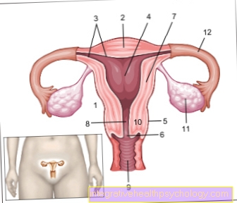
The Rectum lies in the small pelvis. He is very close to the Sacrum (Os sacrum) so more in the back of the pelvis. In women, the front of the rectum borders uterus (Uterus) and the Scabbard (Vagina). In the male it is bordered by the rectum in front Vesicle gland (Glandula vesiculosa) and prostate (Prostate) as well as the Vas deferens (Ductus deferens) and the bladder. The doctor also uses these positional relationships during examinations. In digital rectal examinations, the prostate or uterus can be palpated with the finger over the rectum.
The rectum goes through the pelvic floor by. This is where the transition from rectum to anal canal also lies.
Blood vessels
The rectum is supplied with blood through three larger vessels. The first vessel is the superior rectal artery. This upper rectal artery supplies most of the rectum and the corpus cavernosum recti. This corpus cavernosum recti is an erectile tissue. The erectile tissue becomes filled with blood.During the continence phase or the filling phase of the rectum, the contraction of the two sphincters squeezes the venous outflow of the erectile tissue. This allows the erectile tissue to fill with blood, but not to empty it. This ensures an additional gas-tight seal.
The second vessel for supplying the rectum is the arteria recatlis media. It mainly supplies the lower part of the ampoule. The third vessel is the inferior rectal artery. This supplies the anal canal and the sphincter muscles.
function
To one secure closure of the rectum The rectum and anal canal are equipped with a complex muscle system to allow the stool to be held. This muscle system is also called Sphincter system (Sphincter). The sphincter system is made up of three different muscles.
The internal sphincter (Musculus sphincter ani internus) is a reinforcement of the circular muscle of the rectum. He belongs to smooth muscles and is thus not arbitrarily controllable. The internal sphincter is in constant tension. This muscle only relaxes to defecate.
The external sphincter (Musculus spincter ani externus) clamps the Anal canal from both sides. As a result, the external sphincter forms the anal canal into one narrow slot. The external sphincter is also under constant tension and thus closes the anal canal. In contrast to the internal sphincter, however, the external sphincter is a striated muscle and thus arbitrary controllable.
The last muscle that is counted in the sphincter system is the Puborectalis muscle. This muscle is also striated. The puborectalis muscle surrounds the rectum like a noose. This further increases the bend formed by the perineal flexure. This also contributes to the closure of the rectum. The puborectalis muscle constricts the lumen of the rectum into a slit that is cross-shaped to the other constriction of the external sphincter.
That the stool can be held in the rectum is called Continence designated. The continence is guaranteed by several involved structures. The sphincter system closes the rectum and anal canal in a crossed manner from two sides. In addition, the corpus cavernosum recti fills with blood in the event of a backlog and thus also seals the intestine against any gases that may escape.
Lie in the rectum Stretch and touch receptors. If the rectum now fills with stool, the sensation of an urge to defecate is triggered via these receptors. over Nerve connections the internal sphincter muscle relaxes involuntarily. The external sphincter muscle and the puborectalis muscle also relax. This allows the anal canal to widen, as there is no longer any occlusion of the intestinal lumen. The corpus cavernosum now also empties due to the decreasing muscle tension.
By Longitudinal muscle contractions of the rectum can chair now additionally expelled become. This can be done through the Increase in pressure in the body by means of the Abdominal press still be strengthened and it comes to Emptying (Defecation).
Diseases of the rectum
It can happen that the rectum in one Pelvic floor weakness and the Sphincters falls down. This means that the muscle level here is no longer strong enough to hold the organs. This causes the rectum to collapse on itself and can bulge out through the anus. This incident is also called Rectal prolapse.