Mitochondria
definition
Every body cell has certain functional units, so-called cell organelles. They are the small organs of the cell and, like the large organs, have assigned areas of responsibility. Cell organelles include mitochondria and ribosomes.
The function of the cell organelles are different; some produce building materials, others ensure order and clean up the "rubbish".
Mitochondria are responsible for the energy supply. They have been using the relevant term "power plants of the cell" for many years. In them, all the necessary components for energy generation are brought together in order to produce biological energy suppliers for all processes with what is known as cellular respiration.

Each cell in the body has an average 1000-2000 individual mitochondria, so they make up about a quarter of the entire cell. The more energy a cell needs for its work, the more mitochondria it usually has.
Therefore, nerve and sensory cells, muscle and heart muscle cells are among those that are richer in mitochondria than others, because their processes run almost permanently and are extremely energy-intensive.
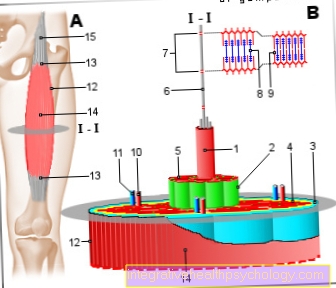
Illustration of mitochondria

- Mitochondria
- Nucleus -
Nucleus - Core body -
Nucleolus - cytoplasm
- Cell membrane -
Plasmallem - Pore canal
- Mitochondrial DNA
- Intermembrane space
- Robisons
- matrix
- Granule
- Inner membrane
- Cristae
- Outer membrane
You can find an overview of all Dr-Gumpert images at: medical illustrations
Structure of a mitochondrion
The structure of a mitochondrion is quite complex compared to other cell organelles. They are about 0.5 µm in size, but can also get larger.
A mitochondrion has two shells, a so-called outer and an inner membrane. The membrane has a size of about 5-7nm.
Read more on the subject at: Cell membrane
These membranes are different. The outer one is oval like a capsule and with its many pores it is permeable for substances. The interior, on the other hand, forms a barrier, but can selectively let substances in and out through many special channels.
Another special feature of the inner membrane compared to the outer membrane is its folding, which ensures that the inner membrane protrudes into the interior of the mitochondrion in innumerable narrow indentations. Thus, the surface of the inner membrane becomes significantly larger than that of the outer one.
This structure creates different spaces within the mitochondrion, which are important for the various steps of energy generation, including the outer membrane, the space between the membranes including the indentations (so-called Christae), the inner membrane and the space within the inner membrane (so-called. Matrix, it is only surrounded by the inner membrane).
Different types of mitochondria
There are three different types of mitochondria known: the saccule type, the cristae type and the tubule type. The division is made on the basis of the invaginations of the inner membrane in the mitochondrial interior. Depending on how these indentations look like, you can determine the type. These folds serve to enlarge the surface (more space for the respiratory chain).
The cristae type has thin, strip-shaped invaginations. The tubular type has tubular invaginations and the saccular type has tubular invaginations that have small bulges.
The Critae type is the most common. The tubular type mainly in cells that produce steroids. The sacculus type is only found in the zona fasciculata of the adrenal cortex.
A fourth type is occasionally mentioned: the prism type. The invaginations of the type appear triangular and it occurs only in special cells (astrocytes) of the liver.
Mitochondrial DNA
In addition to the cell nucleus as the main storage location, mitochondria contain their own DNA. This makes them unique compared to the other cell organelles. Another special feature is that this DNA is in the form of a so-called plasmid and not, as in the cell nucleus, in the form of chromosomes.
This phenomenon can be explained by the so-called endosymbiotic theory, which states that mitochondria were living cells of their own in primeval times. At some point these primordial mitochondria were swallowed by larger unicellular organisms and from then on did their work in the service of the other organism. This cooperation worked so well that the mitochondria lost the properties that characterize them as an independent form of life and have integrated themselves into cell life.
Another argument in favor of this theory is that mitochondria divide and grow independently without needing information from the cell nucleus.
With their DNA, the mitochondria are an exception to the rest of the body, because mitochondrial DNA is strictly inherited from the mother. They are delivered with the mother's egg cell, so to speak, and divide during embryo development until each cell in the body has sufficient mitochondria. Their DNA is identical, which means that maternal lines of inheritance can be traced back for a long time.
Of course, there are also genetic diseases of the mitochondrial DNA, so-called mitochondropathies. However, these can only be passed on from mother to child and are generally extremely rare.
What are the special features of the inheritance of mitochondria?
Mitochondria are a cell compartment that is purely on the maternal side (maternal) is inherited. All children of a mother have the same mitochondrial DNA (abbreviated as mtDNA). This fact can be used in genealogical research by using the mitochondrial DNA, for example, to determine whether a family belongs to a people.
In addition, mitochondria with their mtDNA are not subject to any strict division mechanism, as is the case with DNA within our cell nucleus. While this is doubled and then exactly 50% is transferred to the daughter cell that is created, the mitochondrial DNA is sometimes more and less replicated in the course of the cell cycle and is also distributed unevenly to the newly emerging mitochondria of the daughter cell. The mitochondria usually contain two to ten copies of the mtDNA within their matrix.
The purely maternal origin of the mitochondria can be explained by our germ cells. Since the male sperm only transfers its head, which only contains the DNA from the cell nucleus, when it merges with the egg cell, the maternal egg cell contributes all mitochondria for the development of the later embryo. The tail of the sperm, at the front end of which the mitochondria are located, remains outside the egg, since it only serves the sperm for locomotion.
Function of mitochondria
The term "power plants of the cell" boldly describes the function of the mitochondria, namely energy generation.
All energy sources from food are metabolized here in the last step and converted into chemical or biologically usable energy. The key to this is called ATP (adenosine tri-phosphate), a chemical compound that stores a lot of energy and can release it again through decomposition.
ATP is the universal energy supplier for all processes in all cells, it is needed almost always and everywhere. The last metabolic steps for the utilization of carbohydrates or sugars (so-called cell respiration, see below) and fats (so-called beta-oxidation) take place in the matrix, which means the space inside the mitochondrion.
Proteins are ultimately also used here, but they are already converted into sugars beforehand in the liver and therefore also take the path of cell respiration. Mitochondria are thus the interface for converting food into larger amounts of biologically usable energy.
There are very many mitochondria per cell, roughly you can say that a cell that needs a lot of energy, such as muscle and nerve cells, also has more mitochondria than a cell whose energy expenditure is lower.
Mitochondria can initiate programmed cell death (apoptosis) via the intrinsic signaling pathway (intercellular).
Another task is the storage of calcium.
What is cellular respiration?
Cell respiration is a chemically extremely complex process for converting carbohydrates or fats into ATP, the universal energy carrier, with the help of oxygen.
It is divided into four process units, which in turn consist of a large number of individual chemical reactions: glycolysis, PDH (pyruvate dehydrogenase) reaction, citric acid cycle and respiratory chain.
Glycolysis is the only part of cellular respiration that takes place in the cytoplasm, the rest takes place in the mitochondria. Even small amounts of ATP are produced during glycolysis, so that cells without mitochondria or without oxygen supply can meet their energy needs. However, this type of energy generation is much more inefficient in relation to the sugar used. Two ATP can be obtained from one sugar molecule without mitochondria, with the help of the mitochondria there is a total of 32 ATP.
The structure of the mitochondria is crucial for the further steps of cell respiration. The PDH reaction and citric acid cycle take place in the mitochondrial matrix. The intermediate product of glycolysis is actively transported into the interior of the mitochondrion via transporters in the two membranes, where it can be further processed.
The last step in cell respiration, the respiratory chain, then takes place in the inner membrane and uses the strict separation of the space between the membranes and the matrix. This is where the oxygen we inhale comes into play, which is the last important factor for a functioning energy production.
Read more about this under Cellular respiration in humans
How can mitochondria be strengthened in their function?
Physical and emotional strain can reduce the performance of our mitochondria and thus our body.
You can try to strengthen your mitochondria with simple means. From a medical point of view, this is still controversial, but there are now some studies that ascribe some methods a positive effect.
A balanced diet is also important for mitochondria. A balanced electrolyte balance is particularly relevant. These include above all sodium and potassium, sufficient vitamin B12 and other B vitamins, omega3 fatty acids, iron and the so-called coenzyme Q10, which forms part of the respiratory chain in the inner membrane.
Sufficient exercise and sport stimulates the division and thus the multiplication of mitochondria, as they now have to generate more energy. This is also noticeable in everyday life.
Some studies show that exposure to cold, e.g. taking a cold shower, also promotes division of the mitochondria.
Diets such as a ketogenic diet (avoiding carbohydrates) or intermittent fasting are more controversial. You should always consult your trusted doctor before such measures. Particularly in the case of serious illnesses, such as cancer, one should exercise caution with such experiments. General measures such as exercise and a balanced diet, however, never do any harm and have been shown to strengthen the mitochondria in our body.
Is it possible to multiply mitochondria?
In principle, the organism can regulate the production of mitochondria up or down. The decisive factor for this is the current energy supply of the organ in which the mitochondria are to be multiplied.
A lack of energy within these organ systems ultimately leads to the development of so-called growth factors via a cascade of different proteins that are responsible for registering the lack of energy. The best known is the PGC –1 – α. This in turn ensures that the cells of the organ are stimulated to form more mitochondria in order to counteract the lack of energy, since more mitochondria can also provide more energy.
In practice, this can be achieved, for example, by adjusting the diet. If the body does not have enough carbohydrates or sugar to provide energy, the body switches to other energy sources, such as B. fats and amino acids. However, since their processing is more complicated for the body and energy cannot be made available as quickly, the body reacts by increasing the production of mitochondria.
In summary, we can say that a low-carbohydrate diet or a period of fasting paired with strength training strongly stimulates the formation of new mitochondria in the muscles.
Mitochondrial diseases
Mitochondrial diseases are mostly due to defects in the so-called respiratory chain of the mitochondria. If our tissues are adequately oxygenated, this respiratory chain is responsible for ensuring that the cells here have sufficient energy available to carry out their functions and to keep themselves alive.
Correspondingly, defects in this respiratory chain ultimately result in the death of these cells. This cell death is particularly pronounced in organs or tissues that are dependent on a constant supply of energy. This includes skeletal and cardiac muscles as well as our central nervous system, but also the kidneys and liver.
Those affected usually complain of severe muscle pain after exercise, have reduced mental abilities or may suffer from epileptic seizures. Kidney dysfunction can also occur.
The difficulty for the doctor is to correctly interpret these symptoms. Since not all mitochondria in the body, and sometimes not even all mitochondria in a cell, have this impaired mitochondrial function, the characteristics can vary greatly from person to person. In medicine, however, there are established disease complexes in which several organs are always affected by malfunctions.
- At the Leigh Syndrome For example, cell death in the area of the brain stem and damage to peripheral nerves occur. In the further course, organs such as the heart, liver and kidneys also become susceptible and ultimately stop functioning.
- In the symptom complex of myopathy, encephalopathy, lactic acidosis, stroke-like episodes, briefly MELAS syndrome, the person concerned suffers from cell defects in skeletal muscles and the central nervous system.
These diseases are usually diagnosed using a small tissue sample from a muscle. This tissue sample is examined microscopically for abnormalities. If so-called “ragged red fibers” (a clump of mitochondria) are present, these are a very large indicator of the presence of a mitochondrial disease.
In addition, the components of the respiratory chain are often examined for their function and the mitochondrial DNA is examined for mutations using sequencing.
A treatment or even a cure for mitochondrial diseases is currently (2017) not yet possible.


.jpg)