MRI for optic atrophy
Introduction to optic atrophy MRI
In a study from 2006 to 2007 it was found that MRI can measure the thickness of the optic nerve (nervus opticus).
If there is a loss of nerve fibers (optic atrophy), this becomes visible during the examination in the MRI as a decrease in the diameter of the optic nerve thickness. This method with the 3T-MRT turned out to be an extremely sensitive and non-invasive method, which makes it possible to assess the condition of the optic nerve in a short examination time, especially in glaucoma patients.

General
Magnetic resonance tomography (MRT) is a modern imaging method that is used in ophthalmology in addition to other areas.
The so-called nuclear spin is used during the examination by means of MRI, which means that each atomic nucleus rotates around its own axis and thus becomes a weak magnet. In magnetic resonance tomography, the hydrogen atoms in the body, whose axes otherwise point in different directions, are brought into a parallel arrangement by a strong magnet.
Then radio waves are emitted, which disturb the hydrogen atoms in such a way that their axial directions are changed. After switching off the radio waves, the hydrogen atoms jump back into their original parallel position and send out radio waves themselves, which are then recorded by the MRI machine. A layplan is then calculated from this data for the respective plane.
In principle, only the density of the hydrogen atoms is measured, which means that the anatomical structures in the body and especially the soft tissue can be well represented by the MRI.
MRI for optic atrophy
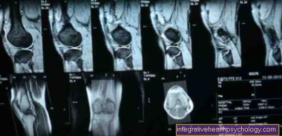
For a precise assessment of structural changes in the eye and eye socket, nuclear spin tomography (magnetic resonance tomography, MRT) is very well suited, as it is a exact differentiation of different tissues and substances is made possible. The procedure is used, among other things, to diagnose Tumors and inflammation applied.
The is then measured Size of structureshow the thickness of the external eye muscles. With inflammation of the muscles or a inflammatory orbital swelling (endocrine orbitopathy, exophthalmos) this can be important and groundbreaking in the diagnosis. Especially with a Inflammation of the optic nerve (Neuritis nervi optici, retrobulbar neuritis), an examination of possible optic atrophy by means of MRI is of particular importance.
How does the MRI work for optic atrophy?
The patient is lying on an examination table in the MRI machine pushed in. The area of the body to be examined, in this case the head for ophthalmology, is brought into position so that it is level with the device.
Then the Sectional images of several Layers of the body recorded, the distance between the layers should be wider than a few millimeters. This can prevent Missing minor changes in tissuethat would otherwise be exactly between two images.
The device gives during the examination loud noises (Click and tap) by yourself, by that Operation of the solenoids originate. For anxious patients, this can be next to the spatial confinement be uncomfortable. It is possible to make one against the noise ear protection to wear. During the examination, the patient is in contact with the examination staff and the The investigation can be canceled if necessary become.
About certain structures more clear Being able to see and assess is partly done before or during the MRI examination Contrast media in a vein given. The examination time of the MRI is up to one half an hourseldom longer, depending on the area of the body to be examined.

