Operation of a hip prosthesis
Synonyms
artificial hip joint, Total hip joint prosthesis (HTEP or HTE), Hip joint prosthesis, total hip endoprosthesis
definition
The designation Total hip joint replacement stands for "artificial hip joint". The artificial hip joint is the human hip joint modeled and thus basically consists of the same parts.
When a hip prosthesis is implanted, the joint socket of the pelvis is replaced by a socket prosthesis (= "artificial socket"). The femoral head and the Femoral neck themselves are replaced by the prosthesis socket with an artificial head.
It is possible to fix the named components in the bone either with or without bone cement.

Appointment with a hip expert?

I would be happy to advise you!
Who am I?
My name is I am a specialist in orthopedics and the founder of .
Various television programs and print media report regularly about my work. On HR television you can see me every 6 weeks live on "Hallo Hessen".
But now enough is indicated ;-)
The hip joint is one of the joints that are exposed to the greatest stress.
The treatment of the hip (e.g. hip arthrosis, hip impingement, etc.) therefore requires a lot of experience.
I treat all hip diseases with a focus on conservative methods.
The aim of any treatment is treatment without surgery.
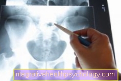
Which therapy achieves the best results in the long term can only be determined after looking at all of the information (Examination, X-ray, ultrasound, MRI, etc.) be assessed.
You can find me in:
- - your orthopedic surgeon
14
Directly to the online appointment arrangement
Unfortunately, it is currently only possible to make an appointment with private health insurers. I hope for your understanding!
Further information about myself can be found at
Therapy / surgery
Prepare for the operation
Since all prosthesis operations involve so-called "Electoral interventions“And the appointment is known over a longer period of time, preparations for the operation can be made early and well thought-out. In addition to obtaining information, preparations include:
- Educational talks with the treating, possibly operating doctor.
- information gathering with regard to the question: Which prosthesis model is suitable for me?
- Obtaining information with regard to the question: Is there Specialists / Specialty clinics?
- There is the possibility of Autologous blood donation?
Course of operation
In short, as part of a hip prosthesis operation, surgically damaged bones or cartilage parts of the hip joint are removed and replaced with artificial parts.
The hip joint consists of the thigh bone (= femur), a long tubular bone that ends with a ball at the top. This "ball" is embedded in the hip socket (= acetabulum) of the pelvis, while ensuring freedom of movement. This construction enables maximum freedom of movement in the form of walking, sitting, ...
Patients for whom a hip joint endoprosthesis has to be considered have lost this maximum freedom of movement or are severely restricted in their ability to carry out everyday movements. The underlying causes in this regard should not be addressed at this point. Rather, it is intended to show how such an operation proceeds.
As already briefly mentioned above, damaged bone or cartilage parts are removed within the framework of the hip endoprosthesis, while trying to preserve healthy tissue. The removed components are replaced by artificial "spare parts". These artificial parts are, on the one hand, the acetabulum, the hip joint socket, the hip shaft with the hip prosthesis head (for examples see above).
The aim of hip prosthesis surgery is to regain maximum quality of life in the form of pain-free movement of the hip joint.
Learn more about: Pain after hip surgery
Access

Every operation requires access to the area to be operated on. In the context of hip arthroplasty, this access can be opened anterolaterally (from the front), laterally (from the side) or posteriorly (from behind).
The size and thus the length of the entrance is individually different and varies between 10 and 30 cm. The surgical team first prepares the area to be operated on, and finally the surgeon cuts through layers of tissue and muscle to allow a free path to the hip joint.
Once this has happened, the femoral head is dislocated from the area of the acetabulum.
Dissection of the femoral head
After opening the operation and dislocating the femoral head from the area of the acetabulum, the femoral head is completely removed. The decisive factor is the height at which the femoral head is severed. This sometimes has a major impact on the course of the operation, but above all on the leg length and thus on the situation after the operation.
Dissection of the acetabulum

The acetabulum must also be prepared. For this purpose - after the acetabulum has been milled out circularly - a shell is inserted into the acetabulum. As already mentioned above, there are different models of such bowls. While so-called press-fit pans are “only” hammered into the pan, there are pans that have to be built in with cement containing antibiotics. To enable undisturbed movement, the diameter of the bowl is usually around 2 mm larger than the diameter of the head. So that the alignment of the shell was not designed incorrectly later, the correct alignment of the shell is checked and, if necessary, corrected during the operation with the help of a target device.
If, in the course of such a check, it is found that the new components appear to be inadequately fixed, this problem can be counteracted in exceptional cases by means of additional screw connections. Under certain circumstances, this can lead to further problems - especially if replacement operations are necessary.
Preparation of the medullary canal

For this purpose, a drill is first used to drill into the medullary canal of the long bone. The use of so-called “rasps” enables the preparation of an area into which the shaft fits exactly. Whether an exact fit is available is first tested before the implant - with or without cement - is installed in the bone.
Choice of femoral head

A femoral head that fits the hip socket is now placed on the shaft. All parts of the prosthesis have thus been implanted. Of course, it is necessary to check the function of the new hip joint before sewing it up.
If possible, it should be possible to rule out that the new hip joint tends to dislocate (= dislocation). It can happen that an artificial hip joint is used dislocation tends. To counteract such cases, “inlays” have been developed that can also be inserted into the cup. They allow a better roofing of the femoral head and can thus prevent the hip joint from dislocating during extreme movements.
Wound closure

After "passing" the function test, the operating area is closed again. This means that the hip joint capsule is first (partially) closed again and any severed muscle parts are anchored again in the area of their origin. Finally, the individual skin layers have to be closed. For this purpose, the surgeon has various suturing techniques or even the option of “tacking together”.
Anesthesia and duration
It must be assumed that a hip joint endoprosthesis operation can take an average of between 45 minutes and 2 hours, with upward and downward deviations being conceivable.
The operation can be carried out under general or partial anesthesia.
At this point it should be pointed out that rehabilitation measures should usually be followed up after the endoprosthetic operation. Which form can be considered for this in individual cases should be discussed with the treating / operating doctor. The motto is: self-help is useful, but too much help, too much ambition can slow down or significantly limit the healing process.
Duration
The duration of the use of a hip prosthesis is made up of or can be divided into:
- Duration of the operation
- Length of hospital stay and
- Duration of the rehabilitation phase afterwards.
1. The operation itself, in which the prosthesis is inserted, takes on average one to one and a half hours from induction of anesthesia to wound closure and anesthesia drainage.
2. After the operation, the patient - provided no complications have arisen - will be cared for in a normal ward for about 7-10 days, whereby the length of stay can often vary due to the postoperative, individual course.
3. Immediately after the hospital stay, there is usually an outpatient or even further inpatient rehabilitation measure, which on average extends over a period of three to four weeks.
Read more about this at: Rehabilitation after installing a hip prosthesis
After about 3 months, the artificial hip joint is usually completely healed and resilient, so that no restrictions are necessary in everyday life.
Anchoring the prosthesis at the top

According to the design of the prosthesis, the hip prosthesis shafts anchor more in the upper part of the prosthesis.
The remaining part of the prosthesis also contributes to the anchorage, but is not as important in percentage terms.
In any case, it is important that the prosthesis shaft is brought as close as possible to the hard part (Compakta) of the tubular bone and that it is accepted by the patient's own bone in the weeks following the prosthesis operation.
This creates a biological - synthetic bond between the prosthesis and the bone, which remains fused with one another for a lifetime.
In particular, a bacterial infection or abrasion particles of the sliding pairing of the femoral head with the acetabulum lead to a loosening of the hip prosthesis.
Denture anchorage below

With this type of prosthesis, the majority of the prosthesis anchorage is in the middle / lower part of the prosthesis. In percentage terms, the upper part of the shaft only contributes less to the anchoring in the femoral bone.
Overall, this type of prosthesis is installed in smaller numbers than the type of prosthesis listed above.
Ultimately, various influencing factors play a role - such as the bone quality - a role in which type of anchorage should be chosen.
The prosthetic models
Which different prosthesis models are there?

The aim is always to restore an undisturbed, painless and, above all, permanent function of the hip joint. Accordingly, a distinction is made between three different types of prosthesis, which differ in the way the prosthesis is anchored in the body's own bone.
These are:
- The cementless prosthesis
- The cemented prosthesis
- The hybrid prosthesis,
which is made up of cemented and uncemented prosthesis parts.
The advantage that there are different options for anchoring the prosthesis in the body's own bone is that the patient can be fitted with a total of three - possibly even more - HTEPs. Despite all the requirements that a prosthetic model has to meet, replacement operations cannot be ruled out and are due within a certain period of time (see below).
In the following, the various types of prostheses are presented and their properties are described.
1. The cementless prosthesis
In contrast to a cemented prosthesis, with a cementless prosthesis the prosthesis shaft and the artificial hip socket are either screwed to the bone or clamped into the bone. In the first case one speaks of a so-called screw socket, in the latter case of a “press-fit prosthesis”.
A fixation of cement-free prostheses, which are usually made of titanium, is achieved in a special way by the special surface coating, which consists of a basic bone substance, the hydroxyapatite. The surrounding bone grows towards the prosthesis, so that a close connection is established between the two substances. Above all, this ensures the direct transmission of the load forces.
2. The cemented prosthesis
Cemented prostheses differ from non-cemented prostheses in that both the acetabulum and the prosthesis shaft are installed with the aid of a fast-hardening, antibiotic-containing bone cement. They therefore do not have a roughened surface that is supposed to cause the ingrowth.
In the case of cemented prostheses, it is important to avoid any gaps that may arise between the cement and the prosthesis and which may be responsible for the loosening of the prosthesis.
3. The hybrid prosthesis
A hybrid prosthesis is a combination of cementless and cemented prosthesis. Either the prosthesis shaft is fixed with the help of a fast-hardening cement, which is usually antibitotic-containing to prevent infections, while the joint socket is anchored without cement, or the other way around.
There are different model variants for all prosthesis types. Determining the right model requires determining the patient's height, weight and bone shape, as well as the demands that he will make on his new hip joint.
As a rule, prior to the operation, the surgeon uses the imaging procedure to draw up a drawing of the hip to be operated on, which he can then use to determine the exact size and model of the hip prosthesis.
Various components of hip arthroplasty are shown below. One recognizes that - depending on the model variants and the manufacturer - there are different models on the market, the advantages and disadvantages of which can only be determined after many years and always depend on individual circumstances.
The acetabulum
The following pictures should show you the differences between cemented and cementless acetabular cups. As mentioned earlier, cementless prostheses always imply one
cementless prostheses imply the use of a socket with a metal alloy (for example, in the form of titanium).
The prosthesis socket
Analogous to the large number of different socket types, there is also a large selection of prosthetic sockets. A distinction is also made here between:
- cemented pans and
- uncemented pans
Especially the uncemented sockets are differentiated again with regard to their main anchorage zones.
The shaft types
Here, too, the manufacturers advertise with different designs. Comparative studies between the various models and their advantages and disadvantages are only carried out to a limited extent. A random selection of different socket models has been compiled below.
Cementless prosthesis

A cementless prosthesis made of titanium is shown. In the area that is inserted into the bone, the prosthesis is roughened so that the boil can establish a connection with the titanium.
An artificial femoral head made of ceramic is attached.
Modular prosthesis system

A modular system, i.e. the stem length can be selected individually depending on the patient and the surgical situation.
Modular systems are often used in the field of exchange operations / exchange operations.
Macro-prosthesis

Another model with a macroporous upper part and a microporous lower part. Macroporous dentures are usually cast so that they are not made of titanium. As a rule, cobalt-chromium-nickel compounds are used for this.
The hip prosthesis head

The femoral head is that part of the entire prosthesis that can be adapted to the situation and individually, or more precisely: has to be adapted. As already described above, this is a modular part of the total prosthesis.
Modular parts of a total prosthesis, be it in the area of the hip prosthesis head or in the area of the shaft types (see above), help to better adapt to individual circumstances. These components enable the surgeon, for example - if it can be considered useful - Leg length differences balance.
There are different materials that are used to manufacture the hip prosthesis heads. Steel alloys or femoral head prostheses made of ceramic are often used.
Advantages and disadvantages can be found with both materials. Hip prosthesis heads made of ceramic are said to be less abrasive, but also more prone to breakage, while steel practically cannot break, but causes more abrasion.
A final assessment of which materials should be categorized as better has not yet been clarified. Research into new materials and improvements to existing materials will certainly continue.
Note autologous donation
In terms of the possibility of Autologous blood donation It should be pointed out at this point that particularly during hip prosthesis surgery, high blood loss can occur. An autologous blood donation then has the advantage that you “donate blood yourself” in advance, just in case.
This is particularly possible due to the fact that this is an elective intervention (see above). The autologous blood donation takes place around two to six weeks before the planned surgery date in the hospital performing the surgery. It has the advantage that the risk of disease transmission via the bloodstream can actually be ruled out, since you get your own blood again. A foreign blood transfusion is also associated with a certain residual risk at all control bodies that have to pass through foreign blood.
A hip prosthesis operation usually leads to a two to three week hospital stay. This is followed by rehabilitative measures, which can be carried out on an outpatient or inpatient basis and which can be designed very differently.
The first mobilization usually takes place on the first day after the operation. It must be pointed out that this must be done under guidance. As a rule, a physiotherapist is responsible for the initial mobilization, who also explains to the patient which movements can be performed and how and which cannot.
X-ray with an artificial hip joint

- Cup of the hip prosthesis
- Prosthetic socket
- Prosthetic head
What should a hip prosthesis be able to do?
Since high demands are placed on hip prostheses and they must meet these requirements in a special way, the requirement profile is that prostheses
- corrosion resistant
- abrasion resistant
- compatible (no allergy)
- Resistant to the pressure and bending loads of body movements
must be. Generally only a few, very specific metal alloys meet this requirement profile. These special alloys include, for example, specific plastics, titanium, ceramics and stainless steel.
Pain during hip replacement surgery
Whether and to what extent pain occurs after the operation to implant the hip joint prosthesis depends on several different factors:
On the one hand, on the type of operation, although now almost without exception the minimally invasive access via an approx. 8-10cm long skin incision on the side of the hip joint is chosen (anterolateral approach).
The advantage here is that neither muscles nor tendons have to be severed on the access path to the hip joint, so that the healing process takes place faster, with fewer complications and with less pain.
In most cases, there is only little pain after the operation, sometimes even no pain at all. With moderate pain medication, most patients are absolutely pain-free after a few hours, but at the latest after 1-2 days - except for a slight wound pain from the scar.
If the pain continues well beyond the second ostoperative day or even gets worse over time, this can be a sign of complications, such as: B. infections, loosening of the prosthesis, calcium deposits in the hip muscles, adhesions or adhesions and a hip dislocation.
Read more on the topic: Hip prosthesis causes pain