Collarbone fracture in the toddler
introduction
The collarbone fracture is one of the most common broken bones in children (approx. 10%). The gender distribution is not completely balanced: boys make up the greater proportion of patients with around 2/3. A broken collarbone can develop in a variety of ways. Most of the fractures can be treated very easily and without surgical intervention.

causes
The causes of a broken collarbone lie in large part in Accident situations. The force can be applied directly to the collarbone, for example if the child falls with the collarbone (Clavicle) hits a solid object, another person or the ground. The direct traumas do about 90% of the broken collarbones. Less common are fractures of the collarbone through indirect violence. This means that the child does not hit the collarbone directly, but absorbs the fall or impact with the hand or elbow. The force that acts on the arm is passed on to the collarbone. Since the collarbone is not built for such enormous forces, it can also fracture (break) in such accidents. This course of the accident can be found e.g. if the child falls over the handlebars on the bicycle and tries to catch himself with his hand or arm.
A broken collarbone can also occur when playing with other children in sports that involve direct physical contact (e.g. soccer). Another cause lies in the birth trauma fracture. At birth the child has to go through the very narrow birth canal get to the outside. It can happen that the child hits the mother's bony structures, e.g. of the Symphysis (the connection of the two pubic bones in the anterior pelvis). If it is a more complicated birth that needs to be helped with the hand or a pair of pliers, then a fracture of the collarbone can also result from this force.
Symptoms
The symptoms in the children turn out to be mild to severe pain in the Shoulder area noticeable. Moving the arm is painful or impossible. The arm is mostly in Relieving posture held on the body.
When moving the arm upwards and against resistance, the child complains of pain and may not be able to perform the movements.
Other symptoms are Swelling in the area of the collarbone, as well as one Redness. Bruising in the shoulder area can also indicate a break.
For fractures with Dislocations (Shifts) show up visible steps in the collarbone, which can also be pushed away if necessary.
Another symptom is when the child lays their head slightly on the injured side, as this reduces the amount of tension on the broken collarbone and reduces pain.
Pain in a collarbone fracture
The pain associated with a clavicle fracture can sometimes very strong be. The periosteum is irritated by the bone fracture, which causes the pain symptoms. Pain relief is already achieved by resting the arm. Because there is no muscle pull on the clavicle, it does not move and the fracture ends are close together so that they can heal. Medicinal can be used in children Pain relief With Paracetamol and Ibuprofen can be achieved. The dosage of the medication must be adapted to the child's body weight, which is why the pediatrician should be consulted with the first dose.
Often this also helps with children Distraction from the pain. Since children are easily distracted by playing or reading, this should be tried often. This reduces the need for pain medication and the child is in better mental health. If the child deals with pain too early, it can become too late Somatization disorders come.
diagnosis
The diagnosis can first be made clinically on the basis of descriptions by the parents and the child about the accident and the localization of the pain
The attending physician can often make a good diagnosis by looking at the child. On the side where the collarbone is broken, the child usually holds the arm in a relieving position. It has the arm placed close to the body and holds the forearm in front of the stomach with the other hand.
Often the shoulder is visibly lower on the affected side.
With fractures (breaks) that are more pronounced, a step formation appears on the collarbone. This results from a tear in the ligament holding apparatus of the collarbone. Normally the clavicle is pulled caudally (below) by these ligaments - if these ligaments rupture or the clavicle breaks, in which the ligaments no longer stabilize the whole bone, the trapezius muscle on the clavicle predominates cranially (above) . This is shown in such a way that the clavicle is dislocated (protruding) upwards and can be pressed caudally (downwards); this is known as the piano key phenomenon.
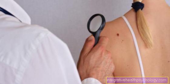
A broken collarbone can usually also be felt by the doctor. The doctor can feel irregularities and fracture gaps through structured palpation of the clavicle.
In the case of fractures in which the skin is spit through the bone, the diagnosis can often be made very quickly. However, a full clinical examination should also be carried out here in order to rule out possible accompanying injuries.
The clinical examination, which should not be neglected, involves palpating and examining neighboring structures (such as the ribs and scapula). Furthermore, it must be absolutely ruled out that no nerves and vessels have been affected.
Ultrasound is the method of choice for further diagnosis. For small children under 2 years of age, this method usually proves to be completely sufficient to show a broken collarbone. For older children and adolescents, it is usually important to carry out an X-ray examination or computed tomography in order to be able to describe the degree of displacement more precisely. During the X-ray, the child holds a weight of around 5 - 10 kg in his hand. By pulling on the arm, a dislocation of the collarbone fracture is better visualized.
In principle, computed tomography should only be performed if the fracture cannot be adequately represented by any other means. Here, too, the rule is that children should be exposed to as little radiation as possible.
Read more on this topic: X-ray examination of the child
Classification of the collarbone fracture
The classification of broken collarbones can be according to Allman in type I, II and III. Further classification options come from Neer, as well as for injuries on Acromioclavicular joint to Rockwood and Dameron.
Allman I describes a break in the middle third of the collarbone. Allman II can be further differentiated by the division of Rockwood and Neer, but basically describes a break in outer third of the collarbone. Allman III describes fractures in the inner third of the collarbone, which ends at the breastbone. A further differentiation can be made here (according to Salter-Harris).
therapy
The following applies to children: bones heal very well. Even severely displaced fractures usually heal by themselves if they repositioned (the fracture surfaces are pushed together).
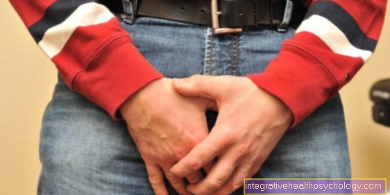
If you have a broken collarbone, surgery is as little as possible. In most cases that is enough Immobilization of the shoulders for a few weeks. For this, the children will be one Backpack bandage created. This is a bandage that is put on like a backpack and pulls the shoulders backwards. This repositions the fracture, i.e. the ends of the fracture are pushed together and fixed. The bandage must be worn for 1-4 weeks, depending on the age of the child. Children under 5 years of age wear it for about 1-2 weeks. Children between 5 and 10 years for about 2-3 weeks. And from the age of 10, 4 weeks of wearing time are indicated.
During this time, the rucksack bandage must be retightened in the first few weeks so that the tension on the fracture point is maintained. It is also important to note that while the bandage is being worn, the Pulse, such as sensitivity and Motor skills be tested on the arms, so that it can be excluded that nerves or blood vessels are pinched off.
In the case of complicated fractures, surgical treatment of the collarbone fracture may be necessary.
In the case of fractures in which nerves and vessels are injured, as well as in open fractures, surgery is required in the majority of cases.In open fractures, the soft tissue structures above the break are severed and there is a gap up to the break.
Often an operation is also performed if the collarbone fracture occurred in the course of an accident in which many other injuries have occurred (multiple trauma). In addition, an operation can be performed if the skin is pierced or if there is a risk of the skin being pierced.
Surgery is also indicated for fractures which, according to radiological imaging, have been classified as fractures of the Rockwood IV-VI type - i.e. fractures in which the fracture is in the outer (lateral) area of the collarbone.
If the conservative treatment, i.e. wearing the rucksack bandage, has not helped (sufficiently), surgery is usually also performed, as there is reason to be concerned that a fake joint (pseudoarthritis) will develop at the incorrectly fused fracture point, which the child will open Long-term pain could cause.
When an operation is performed, various techniques can be used to fix the fracture gap. One possibility is plate osteosynthesis. The fracture is fixed from the outside using a plate that is fixed in both parts of the fracture with nails. However, the method leaves relatively large scars and requires a relatively large surgical access route.
Another method is to fix the fracture with intramedullary Kirschner wires. These are wires that are pushed lengthways through the inner part of the bone, the medullary canal. There are other alternatives with elastic nails, which are intended to reduce the risk of injuries to structures in the area. The advantage of the wires is that a much smaller access path is required for fixation and thus fewer injuries to the tissue are caused.
Backpack bandage
Of the Backpack bandage is placed around both shoulders and fixed on the back. This means that the shoulders are straightened a little and the two fracture parts of the collarbone are placed against each other. Due to the juxtaposition of the fracture parts, the Start the healing process without any obstacles, and the fracture point grows together smoothly. The backpack bandage should be Children up to two weeks, in adults therapy is required for between 3 and 4 weeks.
Prognosis / healing time
In children, a broken bone takes very little time to heal compared to an adult. There are very few known cases in which no healing has taken place.
If the backpack bandage is removed after 1-4 weeks and the child does not show any pressure or movement pain and there are no more instabilities at the break, then it can be assumed that the broken collarbone has healed. A X-ray examination is only available after the bandage has been removed Exceptional cases necessaryif there are still complaints or restrictions.
If an operation has been carried out, no further rest is necessary after removing the fixation. Wires and elastic nails are removed after about 4 -12 weeks; Plates after 4-6 months. Before and after the removal of the wires, plates or nails, an X-ray image is carried out to precisely depict the former fracture gap. Once removed, a sling can be worn for a few days if there is slight pain after the procedure.
Most children have no more problems or complaints after healing. In rare cases, pseudoarthroses (approx. 1%) develop after the operation. These are due to incorrect fixation of the fracture gap, insufficient immobilization of the shoulder or very complicated fractures.
Duration of the collarbone fracture in the child
The collarbone (Clavicle) is in the shoulder. It has two joints in contact with the breastbone (sternum) and to the shoulder joint. The collarbone fracture is a common break in childhood and has healed after about 3-4 weeks. In newborns, a fracture of the collarbone can result from birth trauma. This type of fracture in newborns does not usually require treatment. In small children, the clavicle fracture is the result of external violence. Conservative therapy is usually sufficient here.
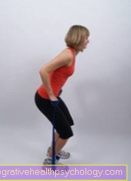
When can my child do sports again?
It takes about 3 to 4 weeks for the collarbone to heal in children. After removing the backpack bandage, physiotherapeutic treatment should be started in order to promote mobility in the shoulder. When in physiotherapy there is no pain and the shoulder moves freely you can slowly start exercising again. In the beginning, you should avoid overloading the shoulder joint and jerky, sweeping movements. If there is pain, the exercise should be stopped.