Visual center
definition
The visual center, also known as the visual cortex, is part of the visual system.
It is located in the occipital lobe of the brain and is part of the central nervous system.
This is where information from nerve fibers in the visual pathway comes in, is processed, interconnected, interpreted and coordinated.
Disturbances in the area of the visual pathway and the visual cortex show up in very different, sometimes characteristic, ways and range from visual field defects to blindness to the inability to recognize faces or objects, for example.
Anatomy & function of the visual center

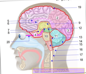
The occipital lobe (Occipital lobe) is the occipital lobe of the brain.
It lies above the cerebellum in the posterior fossa. To the front it borders on the temporal and parietal lobes.
The sulcus calcarinus is an essential landmark in the area of the occipital lobe, in this area lies the visual cortex, also known as the primary and secondary visual cortex.
In order to describe the function of the visual center, the visual pathway upstream of this center must first be briefly discussed, i.e. the path from the eye to the brain.
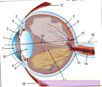
On the way from the eye to the brain, the visual impression passes through several nerve cells. The first nerve cell is in the retina (retina), they are referred to as rods and cones, whereby the rods are primarily used to perceive light, while cones are used to perceive colors.
The second neuron on the way to the brain belongs to the so-called bipolar cells, which are located a little bit in front of the retina in the eye. They pass the impulses on to the ganglion cells, which are also located in the area of the retina. Together with their processes, they form the optic nerve (Optic nerve).
In terms of developmental history, like the retina, this is actually a part of the brain, even if it is "outsourced".
After entering the cranial cavity, the optic nerves on both sides unite to form the so-called chiasma opticum (Optic nerve junction).
This is where all the fibers that cross the outer (lateral or temporal) Map the field of view on the opposite side, which is the inner (medial or nasal) Map the visual field, pull through the chiasm on their original side.
Somewhat confusing is that the lateral field of view is represented on the medial side of the retina and the medial field of view is represented on the lateral side of the retina.
This is due to the fact that the retina is an optical system in which the object that is imaged on it is reduced in size and, above all, the other way around. So again with a camera.
The joins the optic nerve junction Optic tract on.
The left optic tract contains fibers for the visual impression from the left inner (medial) and right outer (lateral) Visual field, the right optic tract fibers from the right nasal and left temporal visual fields.
The optic tract ends in the Corpus geniculatum laterale.
This is in Thalamus. Here the information is switched to the fourth neuron. Before that, some fibers go to the Brain stem ab, these are essential for controlling reflexes.
In everyday life, for example, such a reflex is the coordination of both eyes when looking to the side: If you look to the left with the left eye, the right eye automatically follows.
From Thalamus from the fibers run further than Visual radiation (Radiation optica) to the visual cortex.
The Visual cortex is divided into the primary and they secondary visual cortex.
The primary visual cortex is the first station for the fibers of the visual pathway. It is located in Brodmann-Areal 17 and is also called because of a white stripe that it leaves in the gray matter of the brain Area striata (striped region) designated.
If the impulses come from the eye in the primary visual cortex, what is seen is consciously perceived for the first time, but what is seen is not yet interpreted here.
A certain point corresponds to the Retina a certain area in the cortex, this is called retinotopic structure designated.
The Fovea centralis (Viewing pit), the place of sharpest vision on the retina, takes up 4/5 of the entire primary visual cortex.
The primary visual cortex sends fibers primarily to the secondary visual cortex.
This takes up the Brodmann areas 18 and 19. It wraps around the primary visual cortex like a kind of horseshoe. Here the visual impressions are integrated, analyzed, broken down and interpreted according to size, shape, color, distance and much more.
Nowadays it is known that areas that go beyond the Occipital lobe out into the Temporal and parietal lobes rich, are decisively involved in the secondary processing of visual impulses.
For example, what is seen is linked to what is known so that faces or objects can be recognized.
The secondary visual cortex in turn sends fibers to the Frontal and parietal lobeswhere gaze centers are located that convey, for example, turning the gaze towards or away, corrective movements of the eyes and gaze following movements.
Also pull fibers to Angular gyrus, this is essential for linking what has been seen with language.
Furthermore, fibers from the secondary visual cortex pull into the Brain stem, which is important for reflex movements in the area of the eyes.
clinical understanding of the visual center
Damage to the visual pathway can result from numerous processes:
- Trauma
- Inflammation
- Tumors and others.
Such damage sometimes results in relatively specific loss of vision, depending on where it is located on the visual pathway or the visual system.
A unilateral lesion of the optic nerve leads to unilateral blindness. This can happen, for example, through an optic nerve tear in a traffic accident.
A lesion in the area of the middle part of the optic chiasm leads to a so-called bitemporal hemianopia, which means that the affected person can no longer see anything in the outer field of vision on either side, as the fibers in the chiasm cross in the middle of the opposite side.
Such a failure can occur, for example, due to a tumor in the area of the pituitary gland.
In the area of the brain, a lesion often leads to even more serious failures, as many important processing processes take place here in a small space.
If the primary visual cortex is damaged on one side, this leads - depending on the extent - to minor visual field deficits or to a homonymous hemianopia.
This means that the lateral visual field has failed in one eye and the medial visual field in the other.
This is because the fibers crossing in the chiasm give the left hemisphere fibers, for example, from the medial side of the left visual field and the lateral side of the right visual field.
In the case of processes in the area of the primary visual cortex, the fact that the visual cortex on both sides are very close to one another, however, more often the case that the primary visual cortex on both sides is affected, for example by a tumor in this area.
This can then lead to complete blindness.
Lesions in the area of the secondary visual cortex, on the other hand, do not lead to visual field defects or blindness. In this case, the patient can no longer process and recognize what he has seen. This is known as visual agnosia.
If only a small area of the secondary visual cortex is missing, selective recognition processes can be disturbed, for example only the recognition of faces (Prosopagnosia) to be affected.
The visual system consists of a complex network and switching of fibers on the way from the eye to the brain, where what is seen is only processed to such an extent that it can be consciously perceived and interpreted.