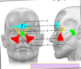
Ethmoid cells
anatomy
The ethmoid gets its name from the ethmoid plate (Lamina cribrosa), which has numerous holes like a sieve and in the facial skull (Viscerocranium) can be found. The ethmoid (Ethmoid bone) is a bony structure between the two eye sockets (Orbits) in the skull. It forms one of the central structures of the sinuses. The interior structure is made of aerated (pneumatized) Ethmoid cells (Cellulae ethmoidalis). The maze of these cells (Labyrinthus ethmoidale) is through bony partitions (Septa) Cut. The ethmoid cells can be divided into anterior and posterior (Cellulae ethmoidales anteriores and Cellulae ethmoidales posteriores) subdivide. The anterior ethmoid cells have a connection to the middle nasal passage (Meatus nasi medius), the posterior to the upper nasal passage (Superior nasal meatus). Some authors differentiate further and also name middle ethmoid cells (Cellulae ethmoidales mediale).

- Frontal sinus -
Frontal sinus - Ethmoid cells -
Cellulae ethmoidales - Maxillary sinus -
Maxillary sinus - Sphenoid sinus -
Sphenoid sinus - Thin septum -
Septum sinuum frontalium
You can find an overview of all Dr-Gumpert images at: medical illustrations
The ethmoid cells boundaries (rear) below to the sphenoid sinuses (Sphenoid sinus), above to the anterior skull base, the Frontal bone (Frontal bone) and to the Ethmoid plate (Lamina cribrosa), on the side are the two Eye sockets, in front the middle ones Corner of the eye (Angulus oculi) and rear middle and front Cranial fossa. Here there is anatomical proximity to the Optic nerve (Optic nerve). Due to the "paper-thin" wall (Lamina papyracea) between the eye sockets and the ethmoid cells can develop Inflammation and tumors spread in both directions. Damage can occur in the area of the thin ethmoid bone plate Inflammation ascends inside the skull.
There are variants regarding the location of ethmoid cells that have proper names. The Haller cells are located in the maxillary sinus and the Onodi cells are located in the sphenoid sinus, where they surround the optic nerve canal (Optic Canal) lie.
Read more about the topic here Sinuses.
Function and tasks
The ethmoid bone stabilizes the bony eye sockets, connects this with the olfactory bulb (Olfactory bulb) and the forehead area and separates cranial cavity and nasal cavity from each other. Together with the nasal septum, it divides the main nasal cavity into two mirror-like areas and thus enables a degree Directional smell. Due to the holes in the ethmoid plate, it is possible that the olfactory threads (Fila olfactoria) and blood vessels (A. ethmoidalis anterior, A. ethmoidalis posterior) can get into the nose to a Blood circulation and sensitivity of the nose to enable. The ethmoid cells also enable the Passage for the nasociliary nerve, a branch of the fifth cranial nerve (trigeminal nerve). This plays an essential role in the Transmission of stimuli between the eyes, upper jaw (Maxilla), Lower jaw (Mandible) and brain. A bony groin, the cockscomb (Crista galli) partially divides the sieve plate and serves as a Attachment of the cerebral sickle (Falx cerebri).
The ethmoid cells participate as the central structure of the paranasal sinuses (Paranasal sinus) at the Air conditioning and thermal insulation of the airways. Through the formation of cavities Saved bones and weight. The anterior ethmoid cells, together with the middle nasal passage and the openings to the maxillary sinuses, form part of a functional unit (Ostiomeatal unit) which to a physiological discharge of secretions contributes. These and other functions and tasks are controversially discussed and are part of it scientific research not yet completed.
Swelling of the ethmoid cells
In the healthy state, there will be mucus Particles and germs through the movement of cells, the stroke of the ciliates, Towards the exit (Ostium, ostiomeatal unit) promoted. As part of a Inflammation of the ethmoid cells (Ethmoid sinusitis) can the Mucous membrane (respiratory ciliated epithelium) of the ethmoid cells swelling. This swelling can lead to the exit (Ostium) and thus the drainage of secretions from the jaw and Frontal sinus (Frontal and maxillary sinuses) to disturb. This also leaves them in the other sinuses Germs and can go to one there further inflammation lead so that a Inflammation and swelling spread to the frontal and maxillary sinuses can.
OP of the ethmoid cells
At a chronic inflammation of the ethmoid cells and adjacent structures are tried through a Clearance to ensure a better drainage of secretions again. The entire system of the paranasal sinus is not cleared out, but only swollen mucous membrane and polyps, as well as the thin bony walls between the ethmoid cells are removed. This is carried out as an endonasal procedure, i.e. it is only operated inside the nose, without any external cut. The operation takes place under general anesthesia and the Hospitalization usually lasts a week. There follows one Aftercare, to keep the nasal passages open. This can last three months or more. After the operation there is usually no longer any swelling, redness or other signs of inflammation, but they can a headache occur.
A detailed explanation by the surgeon and the anesthetist takes place before the operation.
Tumor of the ethmoid cells
A distinction is made between benign (benign) of malicious (malignant) Tumors. Benign tumors in the sinuses are mostly Bone tumors (Osteomas) or invaded Wart growths (infiltrating papillomas).
causes
Tumors of the ethmoid cells can be caused by environmental factors such as Wood dust, chemical fumes or smoke and are recognized as occupational diseases, e.g. among carpenters. Also genetic factors cannot be completely ruled out and are discussed.
Symptoms

Early symptoms of a malignant tumor of the ethmoid cells or the other sinuses can include unilateral nasal obstruction, inflammation symptoms of the ethmoid cells (swelling, redness, pain, pus) and repetitive, frequent nosebleeds (Epistaxis) be. Later on, the cheek, eyelids, and forehead may swell. Double vision disturbances can also occur if the eyeball is displaced by the pressure.
Read more about the topic here: Causes of nosebleeds
diagnosis
First there is a Nasoscopy (Rhinoscopy) to identify a possible tumor directly. By imaging tests, like X-rays, the Computed tomography (CT) and Magnetic resonance imaging (MRI) the extent of the tumor can be recorded more precisely. Also is a comprehensive one Palpation of the cervical lymph nodes absolutely necessary.
therapy
Mostly becomes one surgical removal of the tumor is advisable. In the case of larger malignant tumors, a Radiation and chemotherapy carried out. Small benign osteomas usually do not require any therapy. However, papillomas grow quickly and partially contain also malicious parts. So you should exactly how malignant tumors are treated.
forecast
The The prognosis varies depending on the type of tumor, but is usually quite good if detected in the early stages. At Burglary into surrounding structures such as the eye socket and the palatal fossa (Pterygopalatine fossa), however, the prognosis is mostly comparatively bad.
Inflammation of the ethmoid cells
One distinguishes one depending on the length of the complaints acute (2 weeks duration) of one subacute (more than 2 weeks, less than 2 months duration) of one chronic (over 2 months duration) Inflammation of the ethmoid cells (Sinusitis). The ethmoid cells are the only sinuses that are largely present in their complete structure at birth. Hence one shows up Sinusitis in children mostly in the ethmoid area and in adults more in the maxillary sinus area.
Read more about the topic here: Inflammation of the ethmoid cells
causes
There is inflammation of the ethmoid cells mostly the result of an inflammation of the nasal mucous membrane (Rhinitis or rinosinusitis), but can also be done by a Tooth root disease arise. Further causes and favorable factors can e.g. Nasal packing, closure of the choanas (Chonal atresia), Nasal polyps (Polyposis nasi), Tumors, Resistance and immune deficiency, foreign body, Cystic fibrosis and Damage to the mucus cleansing (mucociliary clearance) be through nasal drops. Often there is bacterial inflammation. Often it is a Mixed infection. A purulent odor suggests an underlying Dental disease down. In rare cases you can also Mushrooms be causative.
Symptoms and Diagnosis
These are characteristic of inflammation of the ethmoid cells Pus streets in the middle nasal passage, Pain, Print- and Knock sensitivity side of the nose and decreased ability to smell (Hyposomy). Radiological imaging (x-rays and computed tomography (CT)) are used for further diagnosis. These show with chronic ethmoid cell inflammation usually bilateral shading.
therapy
With acute sinusitis are mostly therapeutic decongestant nasal drops, Beta-lactam antibiotics and high nose pads recommended.
What is going on when the ethmoid cells are shaded?
When a Inflammation of the ethmoid cells or other sinuses at least twice a year occur, one speaks of a recurrent acute sinusitis. When on computed tomography (CT) shading on both sides of the ethmoid cells persist, this may be an indication of a chronic inflammation of the ethmoid cells be. Unilateral shading can also affect one benign (benign) tumor Clues.
Pain in the ethmoid cells
As part of a Inflammation of the ethmoid cells (Sinusitis) it can be too strong Pain come in the sinuses. This pain can be triggered and aggravated by bending over, coughing or knocking, i.e. in situations where the pressure is increased. In addition, especially if the maxillary sinuses are also affected, tapping and tenderness on the side of the nose can occur. Often shine this pain in the too upper jaw, the teeth and the skin between the upper lip and lower eyelid.


.jpg)