Vena Cava
Synonyms
Vena cava
English: vena cava
definition
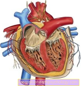
At the vena cava (Vena cava) it is a large blood vessel that has the task of collecting blood in the body and returning it to the heart. A distinction is made between an upper and a lower part. The vena cava opens into the right atrium.
Classification
The vena cava is divided into two sections:
- superior cavernous vein (Superior vena cava)
- inferior vena cava (Inferior vena cava)
Anatomy of the superior vena cava and its tributaries

The superior vena cava (Superior vena cava) runs in the chest (thorax) to the right of the midline along the right edge of the sternum (Sternum). It arises at the level of the 1st rib (ribs) by the confluence of the veins, which the oxygen-poor blood from Arms, head and neck bring about. From behind it borders on the right main bronchus (lung) of the airways. In addition, the azygos vein joins the superior vena cava at the level of the third rib. This is a venous system on the posterior wall of the chest, which, among other things, drains the blood of the esophagus, pericardium, upper diaphragm and bronchi. The hemiazygos vein also flows into the azygos vein. The two are cavocaval anastomoses. This means that they connect the lower and upper vena cava and can be viewed as a bypass circuit in the event of a blood flow disorder in the vena cava.
Anatomy of the inferior vena cava and its tributaries
The inferior vena cava (Inferior vena cava) arises from the confluence of the two pelvic veins (Common iliac veins). It runs from the 5th lumbar vertebrae (lumbar spine) to the right of the main artery (aorta) upwards. The blood flows from the unpaired abdominal organs (e.g. intestines) via the portal vein (Port vein) and thus via the liver and only then shortly before it passes through the diaphragm into the inferior vena cava. The venous blood of the other pelvic and abdominal organs flows directly through the inferior vena cava. After passing through the diaphragmatic hole (Foramen venae cavae) it runs about 1 cm in the chest and then flows into the right atrium together with the superior vena cava.
As a direct tributary it receives the lower diaphragmatic veins (Inferior phrenic veins), Lumbar veins (Lumbar veins), Hepatic veins (Hepatic veins), Renal veins (Renal vein) as well as testicles or ovaries (Vena testicularis or Venae ovariacae).
function
The vena cava (Vena cava) has the task of collecting the blood from the periphery of the body and returning it to the heart. It is also jointly responsible for the filling of the right heart. The pressure in the vena cava is between 0 and 15 mmHg. The pressure shows breathing-dependent and pulse-synchronous fluctuations, which is known as venous pulse. This pressure can be determined and, as a diagnostic measure, especially in intensive therapy, it can be important for assessing the cardiovascular function. This central venous pressure depends on the blood filling of the circulatory system and the pumping capacity of the heart. It is also dependent on the suction effect of breathing, the valve level mechanism of the heart's action, the arteriovenous pressure gradient and the pumping force of the heart.
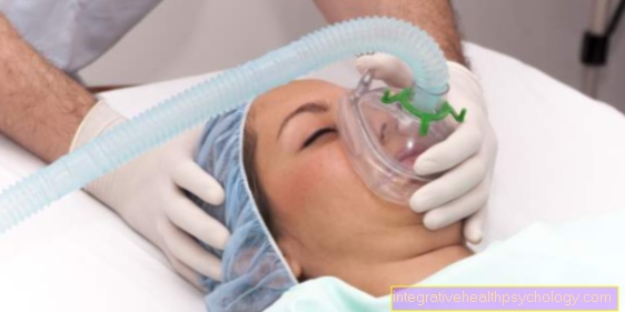
The suction effect through breathing arises because the pressure in the chest drops to negative pressure values during inhalation. This is how blood is sucked in from the periphery. At the same time, the lowering of the diaphragm when you inhale increases the pressure in the abdomen, which causes the abdominal vessels to be compressed, which in turn increases the return flow to the heart.
Heart valves function like valves that only allow blood to pass in one direction. In the heart, all heart valves are in one plane. During the heart's action, the shortening of the muscle fibers shifts this valve level and thus creates an additional suction to support the venous return flow.
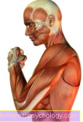
The skeletal muscles shorten and thicken when activated. The thickening compresses and squeezes the veins in the legs. Venous valves in the leg veins prevent the blood from sinking back into the legs. The vena cava itself has no venous valves.
Histological structure
The Vein walls I am subdivided into layers. The layers are thinner than the walls of the arteries. The Intima, a single layer of specialized cells (Endothelial cells). The endothelial cell layer closes the Internal elastic membrane, a braid of elastic fibers drawn from the smooth muscle cells of the subsequent Media to be produced. It follows the External elastic membrane. The last layer is the Adventitia. It consists of connective tissue and anchors a vessel in the area. In the vena cava, the adventitia contains smooth muscle fibers arranged lengthways. Blood vessels must also be supplied by small blood vessels (Vasa vasorum), which run in the adventitia and can penetrate to the media.
Diagnostics and therapy
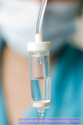
A catheter that goes into the vena cava (Vena cava) is introduced to the right atrium, can be used in diagnostics to assess the cardiovascular function (cardiovascular system). In addition to measuring this central venous pressure (ZVD) the catheter is also used for infusion therapy, which supports infusion therapy with peripheral indwelling venous cannulas. Certain drugs may only be used through a CVC (central venous catheter) can be administered. Artificial nutrition is also possible. The installation of such a central venous catheter must always be absolutely sterile. Because of the risk of infection, a CVC should never be applied longer than necessary and must be changed regularly if it is used for a longer period of time.