Check-ups during pregnancy
Synonyms in a broader sense
Pregnancy care, controls during pregnancy
definition

Preventive care during pregnancy includes regular examinations and consultations for the pregnant woman throughout the pregnancy.
The task of this continuous care is to detect signs of complications and disorders as early as possible, to notice high-risk births and pregnancies and to take appropriate action. Both maternal and child health are the focus of the examinations.
Read more on the topic: Pregnancy complications
The preventive medical checkups during pregnancy have been shown to reduce the mortality of mother and child as well as their diseases. The routine program of preventive medical checkups during pregnancy is set out in the maternity guidelines. The results of these examinations are entered and noted in the so-called maternity record, so that communication between doctors, midwives and the clinic is simplified.
According to the guidelines, the examinations should initially be carried out once a month. Twice a month in the last two months of pregnancy.
The examination in early pregnancy
The first examination with a detailed consultation should take place before a pregnancy, especially for couples with known family diseases, so that this can be well planned and nothing stands in the way of a normal course. During such an examination, diseases of the mother such as Diabetes (Diabetes mellitus) and malformations of the birth canal are identified and discussed at an early stage.
But also lifestyles that are dangerous for the future child, such as Smoke, alcohol or drug consumption should be discovered in such an investigation and a solution to avoid these substances should be found together. This advisory examination thus serves in particular to prevent risks and to treat them before the start of pregnancy.
The first examination after the fertilization should as soon as possible after the absence of the Menstrual bleeding (menstruation) respectively. it includes
- the diagnosis of pregnancy,
- ascertaining the history of the pregnant woman,
- the provisions of the due date,
- physical and gynecological examination,
- Ultrasound examination and
- a detailed consultation
Read more about this topic under Gynecological examination
1. Diagnosis of pregnancy
The Diagnosis of pregnancy is done by specifying the missed menstrual period (secondary amenorrhea). The diagnosis of early pregnancy is now supported by an Ultrasound examination (Sonography) and, if necessary, by an additional hormone test, which proves the hormone HCG that the placenta (placenta) is formed in detectable quantities at a very early stage in pregnancy.
Earlier when the above methods were not yet available for diagnosis, the diagnosis was based on Pregnancy signswhich have been divided into certain, probable and unsafe. The sure signs of pregnancy include hearing from infantile Heart sounds and the feeling of child's body parts as well as child's movements. The mother's physical appearance is considered a likely sign of pregnancy.
These are the lack of menstrual bleeding, the increasing color (pigmentation) the nipple and the vagina as well as the changes in chest and uterus. The increase in size and the changed texture (consistency) of the uterus represent a very important examination option for diagnosis, which is still used today.
The doctor can feel loosening, expansion and pulsating vessels by palpating the uterus and confirm an existing pregnancy. Indigestion, especially vomiting and nausea, frequent urination and changes in mental mood are among the uSafe signs of pregnancy.
2. Survey of the history
In order to ascertain the previous history of the pregnant woman, questions are asked about previous illnesses, especially about possible illnesses after fertilization.
You can find more information on this here: Infections in pregnancy
It is particularly important to include drugs that have been taken for treatment. Because some pathogens and drugs can get into the child's body via the placenta and endanger the child.
Please also read our topic: Medication in Pregnancy
In order to record the physical changes caused by the pregnancy, the doctor asks the pregnant woman about the current state of health. Previous births and pregnancies will also be asked about in this conversation. The manner of previous deliveries, the length of pregnancy and the time after delivery are also important information for the doctor. This first intensive discussion is an important step in the care of the pregnancy and the cooperation between the pregnant woman and the treating doctor.
Learn more about the changes in the body during pregnancy at: Pregnancy symptoms
3. Due date
To the Determine the due date, questions must first be asked about the woman's most recent menstrual periods and cycle. The regularity, the duration and the interval between two bleeds play an important role.
Also the date of last menstrual period and their duration and strength are important for calculating the due date. If the last bleeding that occurred was weaker or shorter than usual, there could also have been so-called implantation bleeding, which occurs shortly after fertilization and determines the time of implantation in the uterine wall. This date would result in a too late due date for the calculation, since a too late start of pregnancy is simulated. If the date of fertilization is known, this is also documented.
The first way to determine the due date is the day of fertilization plus 267 days with a fluctuation of about 7 days. Since the date of fertilization is rarely known, there is another way of calculating it based on the information on the monthly cycle. The so-called Naegele's rule. It uses the first day of the last menstrual period and the interval between two menstrual periods as a basis. One speaks of a shortened cycle if the interval is less than 28 days and of a longer one if the interval is more than 28 days.
Nagele's rule
The Nagele rule is now as follows:
1st day of the last period plus one year plus 7 days minus 3 months plus / minus the deviation from a 28-day cycle.
This calculation, which seems complicated, becomes much clearer with an example.
If the 1st day of a woman's last period was 08/17/2008 and the extended cycle of this woman is 32 days, then 08/17/2008 + one year = 08/17/2009 - 7 days = 08/10/2009 - 3 months = 05/10 .2008 + 4 days deviation from the 28-day cycle = 14.05.2009.
If the woman has a shortened cycle of 24 days, these are deducted so that the result is: 08/17/2008 + one year = 08/17/2009 - 7 days = 08/10/2009 - 3 months = 05/10/2008 - 4 days deviation from the 28-day cycle = 05/06/2009.
It should be noted that this calculation not exactly is to be adopted. Two-thirds of all children are born within 3 weeks of the due date determined using this calculation and only 3.9% of children are actually born on the calculated day.
The due date determined with this invoice therefore only represents one Guideline and in no way represents an absolute value.
Especially in early pregnancy it is possible to measure the child with the help of Ultrasound imaging a fairly accurate statement of the child's age and due date.
The length from the top of the head to the rump of the child is measured as well as the diameter of the Amniotic sac and the child's head. The dimensions determined are then compared with a standard curve. The results of the examinations are documented in the maternity record and the previously calculated due date is adjusted to these results if necessary.
However, these examinations are only meaningful up to the 12th week of pregnancy, since the development of the children is very different at later times.
4. Physical examination
The physical examination at the beginning of pregnancy includes, in particular, the determination of the mother's body weight in order to monitor the progress of the Weight gain to be able to determine in the further course of pregnancy.
Likewise belong one Urinalysis and the Blood pressure measurement for example, to detect pregnancy poisoning at an early stage. In addition, the maternal blood group is determined, the iron content of the blood is determined and the blood of the pregnant woman is examined for infectious agents and antibodies.
A HIV test is only carried out with the consent of the pregnant woman and only the execution of the examination but not the result is documented in the maternity record. All other results are entered in the mother's passport.
As part of an addiction test (First trimester screening) two hormones are determined in the pregnant woman's blood. These are the free HCG, which is produced by the placenta, and the hormone PAPP-A, which is specific for pregnancy. The determination of the concentrations can be used to detect a chromosome-related disease in the child, taking into account ultrasound results. These become noticeable in a changed number of chromosomes in all of the child's body cells. Probably the most well-known disease with a chromosome-related cause is that Down syndrom.
The so-called Triple test the presence of a child's chromosomal disorder, particularly Down's syndrome. This test determines three levels of hormones and is done around the 16th week of pregnancy. However, the results obtained are not always accurate and must definitely be clarified through further investigations.
The gynecological examination includes palpation of the uterus as well as the Ovaries. In addition, cells are removed from the cervix taken.
5. Ultrasound screening examinations
According to the maternity guidelines, three preventive examinations with ultrasound take place during pregnancy.
These should be done something in the 10th, 20th and 30th week of pregnancy.
In addition to determining the due date and age of the child, as mentioned above, the first ultrasound examination serves to monitor pregnancy and early detection of childhood diseases. This examines whether the fertilized egg has properly implanted itself in the uterus, but also whether there is a multiple pregnancy.
In addition, the transparency of the neck is measured and it is checked whether water has accumulated in the area of the child's neck and forms a so-called dorsonuchal edema there. These findings can point to Down syndrome and to heart or kidney disease in the child.
Read more on the topic: Ultrasound examination during pregnancy or placental insufficiency
6. Advice
The very first examination of the pregnant woman should be accompanied by a detailed one consultation with recommendations and instructions on how to behave during pregnancy. These are briefly outlined below, but in no way replace such advice.
Through the nutrition the mother must be supplied with enough nutrients so that both mother and child have sufficient energy. With the common diet in Germany, you do not have to eat for two people, but the usual amount of food is completely sufficient.
A balanced diet is important. It is also based on the increased need Proteins to think these are mainly found in eggs, meat, and fish.
The increased need for Minerals and Trace elementsn, which iodine, iron and Folic acid concerns and can possibly be administered through appropriate tablets. The increased consumption of milk and dairy products increases the need for calcium and magnesium covered.
Of the Use of alcohol, smoking and other drugs should be completely discontinued, as this leads to disorders of the child's development up to malformations and complications of childbirth. Passive smoking should also be avoided consistently. Self Medication can impair child development and should therefore only be taken if there are important reasons and only after medical advice.
If the pregnancy is normal it can be easy Sportstypes like swim or hiking have a positive effect. However, competitive sports and all sports with strong vibrations or an increased risk of falling, such as skiing or athletics, should be avoided. Hard physical work is also to be avoided in the household or at work.
Short to travel, especially in countries with no climatic pollution due to fluctuations in temperatures or altitudes, are also possible during pregnancy. In the middle of pregnancy, this is associated with the lowest risk. To be on the safe side, a check-up should be carried out by the doctor before starting the trip.
In general, rapid temperature changes and excessively high temperatures should be avoided. This also applies during the have a shower or that to bathe to be observed. Sauna sessions longer than 10 minutes should also be avoided.
If the pregnancy is normal and no cervical dysfunction has been detected, there are no restrictions Intercourse necessary. However, it is recommended to restrain sexual intercourse until the beginning of the third month of pregnancy and in the last month before the birth, as this can lead to premature rupture of the bladder or labor.
Further examinations during pregnancy
The physical examinations hardly differ from those already mentioned. They also include weight and blood pressure measurements and the examination of urine and blood. The gynecological examinations such as the assessment of the uterus takes place on the gynecological chair as usual until around the 16th week of pregnancy.
Then it is performed on the couch. Here, too, the vaginal examination is important, in which cells are taken for assessment and the cervix and the cervix (Cervix) be assessed.
Around the 24th week of pregnancy becomes the pregnant woman so-called oral glucose tolerance test is recommended, which is a possible diabetes (Diabetes mellitus) reveals. Such a disease can only appear and be recognized during pregnancy due to the changed hormone concentration, and then it is recognized Gestational diabetes (gastric diabetes) called.
The child is also observed during the preventive medical check-ups. You can do the child's heart work with the help of the Ultrasound Make visible from about the 5th week of pregnancy. The children's movements can also be seen here. The first-time mother can feel this herself from the 20th week of pregnancy.
Mothers who have already given birth to several children notice these movements a few weeks earlier. It is important that the pregnant woman knows that child movements that decrease or change must be clarified by the doctor. The child's movements can also be recorded objectively. The so-called Kineto cardiotocography (K-CTG).
With the help of the ultrasound device, statements are made about the development of the child and its condition in the 20th week of pregnancy. Various malfunctions can be excluded or abnormalities can be investigated at an early stage. The amniotic fluid as well as the Plaster cake (placenta), are also assessed and provide important insights into the care of the child.
Examination at the end of pregnancy

The investigations and deliberations towards the end of the pregnancy serve to prepare for the upcoming birth.
Read more about this at: Last trimester of pregnancy
During this time, the pregnant woman should decide on a maternity hospital and introduce herself to it.
This makes the later birth process much easier, as the circumstances and the personnel can be learned at an early stage and the possibilities of delivery can be discussed.
Important findings that are gained with the help of the third ultrasound examination are important points for the birth, such as the position of the mother cake and that of the child.
It is important that the pregnant woman knows the onset of regular Labor pains as well as the departure of Amniotic fluid around the calculated due date should absolutely lead to her going to the maternity hospital immediately.
In addition, a Birth preparation course that contains information about the birth and care of the newborn.
Participation in the Pregnancy gymnastics is noticeable during the birth through the breathing, positioning and relaxation exercises learned there in a lower need for painkillers and a faster birth process.
Especially around the due date and especially when it is exceeded, a close control of the child's is required Heart sounds and uterine activity with the help of Cardiotocography (CTG) needed to give the doctor a picture of the child's condition.
If the due date is exceeded, appropriate examinations are carried out every 2 days in order to be adequately informed about the maternal and child's condition.
Read here which CTG values are normal!
Read more about this topic under Gynecological examination
Further options for assessing child health
In addition to the methods mentioned above, there are other options available. These are especially offered to pregnant women over 35 years of age. What these methods have in common is that different child cells are removed in order to make their own Chromosomes to investigate. In addition, this method harbors certain risks due to the procedure, which also include the risk of miscarriage.
For this reason, a detailed discussion with the attending doctor takes place before such an operation. The following investigations are important methods:
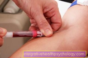
The Amniocentesis (Amniotic fluid examination), in which amniotic fluid is taken through the maternal abdominal wall, usually takes place between the 15th and 18th week of pregnancy. In the removed amniotic fluid there are childish cells that can be examined for different clinical pictures. Another possibility is that Chorionic villus sampling Here, a tissue sample is taken from part of the mother cake and also analyzed. The removal can take place through the vagina as well as through the abdominal wall. In the Fetal blood collection some child's blood is taken from the umbilical cord under ultrasound, which is examined for possible diseases.
The low-risk variant of determining a chromosomal disorder is now that of des Prenatal testswhere only blood is drawn from the mother.
In addition, various optical devices allow a view of the amniotic fluid at the end of the pregnancy, what is called Amnioscopy is referred to, or a direct view of the child, which Fetoscopy is called.