appendix
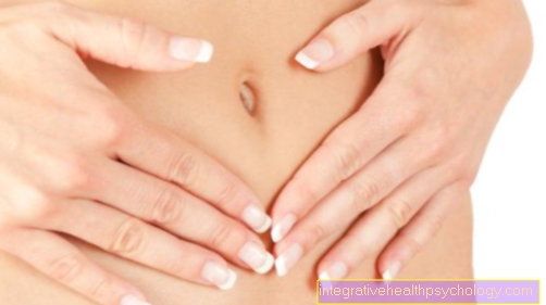
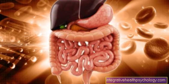
The cecum is part of the large intestine and joins the small intestine. It forms the starting part of the appendix, so to speak. The appendix includes what is known as the appendix vermiformis. That's also the real part of a
The cecum is part of the large intestine and joins the small intestine. It forms the starting part of the appendix, so to speak. The appendix includes what is known as the appendix vermiformis. That's also the real part of a
The connective tissue, cord-shaped structures that surround the uterus on all sides and stabilize it in the pelvis are referred to as uterine ligaments. During pregnancy, the ligaments must expand with the uterus, which is often painful
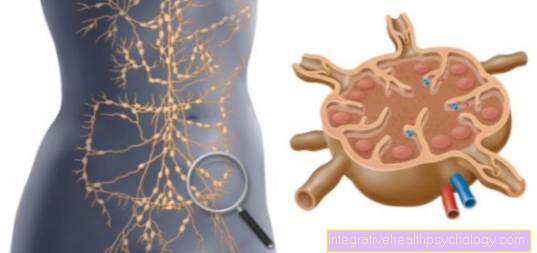
Lymph nodes are small, approximately 2mm wide, kidney-shaped collecting stations in which the lymph is filtered out of the tissue and checked for pathogens. Lymphocytes eliminate the pathogen in the lymph nodes and thus prevent them from becoming infected
The stretching (also "extension") is possible on hinge and ball joints and the opposite form of movement to flexion.
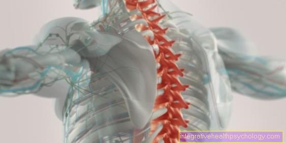
The spinal canal is also called the spinal canal or spinal canal. It is formed by the foramina vertebralis of the vertebral bodies of the cervical, thoracic and lumbar spine as well as the sacrum. It forms the bony structure for the spinal cord, the
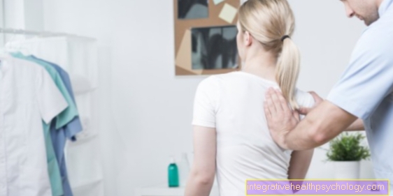
A bursa is a fluid-filled sac that is often found between joints. Its main function is to relieve the pressure and friction that occurs between two bones of a joint when moving, and thus the bones
The joint capsule surrounds a joint and seals off the mechanical movement of the articulating bones towards the outside. The outer layer consists of connective tissue, which gives the capsule a certain strength. The inner layer of the capsule represents
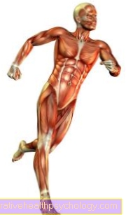
Tendons are the connecting pieces between muscles and bones and should not be confused with ligaments. The most famous tendons include the Achilles tendon, quadriceps tendon, patellar tendon, biceps tendon, triceps tendon, and the rotator cuff. At sportier
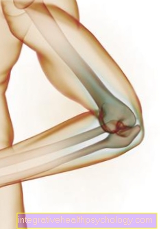
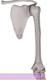
The humerus connects the shoulder joint with the elbow joint and thus with the forearm. At the shoulder joint, the humerus and the shoulder blade (scapula) form a so-called ball joint. At the elbow joint, the humerus forms with the
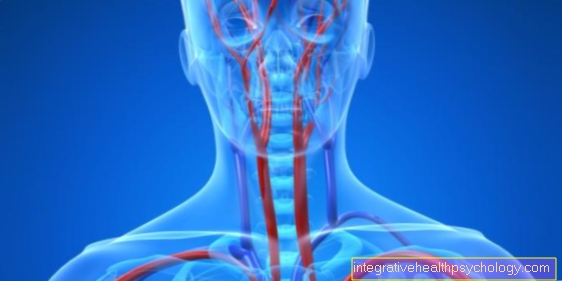
The vertebral artery runs in pairs up the spine and unites to form the basilar artery. Your blood supplies parts of the brain and spinal cord with oxygen. When the artery is narrowed, it is called the vertebral artery synroma
The sciatic nerve is the longest, largest, and thickest peripheral nerve in humans. It comes from a plexus of nerves, the lumbar sacrum nerve plexus (plexus lumbosacralis), which supplies the leg and the buttocks region. When there is damage to the nerve
The tailbone is made up of about four to five individual vertebrae. However, these vertebrae are fused into a single unified bone through what is known as synostosis. The coccyx represents the lower (caudal) end of the vertebra
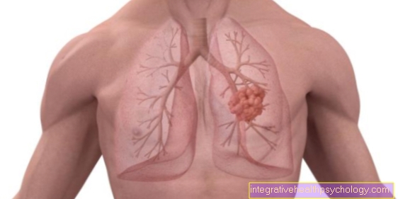
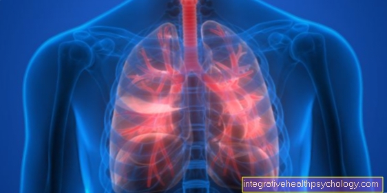
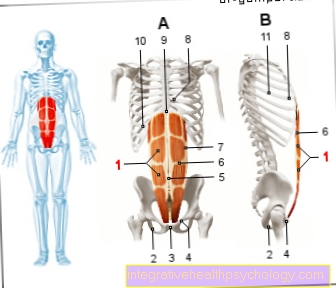
The respiratory muscles are grouped around the lungs and facilitate their movement when breathing. The most important respiratory muscle is the diaphragm. It separates the chest from the abdomen. The muscles that attach to the rib cage are auxiliary respiratory muscles that are used in the
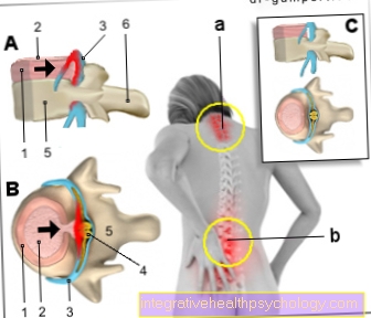
the medical table of figures. Here you will find helpful images on the subject of a slipped disc
The flexion (also "flexion") is possible on ball and hinge joints and represents the opposite form of movement to extension.
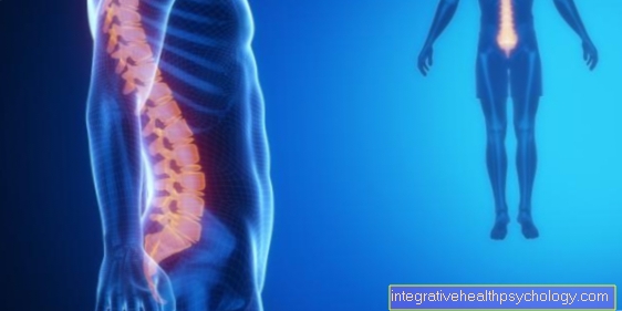
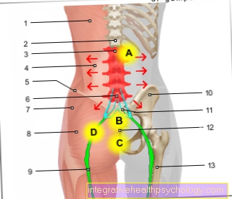
the medical table of figures. Here you will find helpful images on the subject of lumbar spine syndrome.
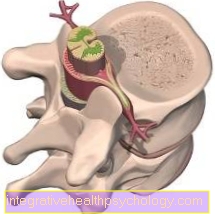
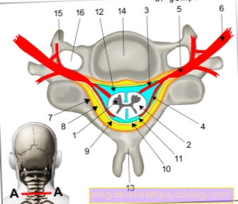
the medical table of figures. Here you will find helpful illustrations on the subject of the spinal cord.
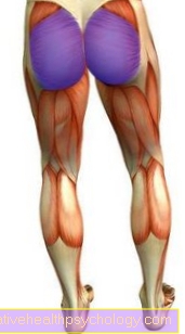
The gluteus maximus (gluteus maximus muscle) covers the buttocks area and is responsible for stretching the thighbone in the hip backwards. In addition, it prevents tipping over when the upper body is bent forward through strong contraction
The pear-shaped muscle (M. piriformis) belongs to the deep hip muscles and connects the sacrum and the thigh bone from behind. Its functions include external rotation and the splaying of the thigh. In its immediate
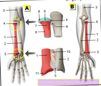
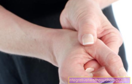
The spoke (radius) and the ulna form the bones of the forearm. Together with the carpal bones, the lunar bone and the scaphoid bone, the spoke forms the essential part of the wrist. Towards the elbow the spoke narrows and ends with the radius k