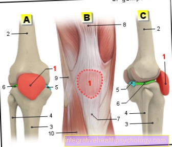
Figure meniscus

- Inner meniscus -
Meniscus medialis - Inner articular knot
(Shinb.) -
Medial condyle - Transverse ligament of the knee joint -
Lig. Transversum genus - Kneecap ligament -
Ligamentum patellae - Bursa - Bursa
- Outer meniscus -
Lateral meniscus - Outer joint nodules
(Shinb.) -
Lateral condyle - Anterior cruciate ligament -
Lig. Cruciatum anterius - Posterior cruciate ligament -
Ligamentum cruciatum posterius - Femur - Femur
- Shin - Tibia
- Kneecap - patella
You can find an overview of all Dr-Gumpert images at: medical illustrations
Related images

Illustration
posterior cruciate ligament

Illustration
Torn knee ligament

Illustration
Inner knee ligament tear

Illustration
Kneecap

Illustration
Outside knee pain

Illustration
Inside knee pain

Illustration
Cruciate ligament

Illustration
Meniscal tear

Illustration
Shin

Illustration
Fibula


.jpg)