Arthrofibrosis in the knee
Synonyms in a broader sense
- Joint scarring
- intra-articular scarring
- "Painful restriction of movement in the knee"
- Cyclops Syndrome
- infrapatellar contracture syndrome / patella baja
- generalized inflammatory joint reaction
definition
The Arthrofibrosis is a dreaded joint disease, largely unexplained in its etiology, after surgical interventions or injuries, which results in a more or less severe and sometimes painful limitation of joint mobility.
A distinction is made between:
- Primary arthrofibrosiswhich is characterized by generalized scarring in the joint.
- Secondary arthrofibrosis, in which local mechanical irritators are the cause of restricted mobility.
Most of the studies in the literature dealt with the development of arthrofibrosis of the knee joint after injuries and Cruciate ligament surgery.
From a clinical point of view, arthrofibrosis of the Knee joint due to a permanent restriction of movement of > 10° for the Elongation and <125° for the diffraction Are defined.
Symptoms
Characteristic of arthrofibrosis is the restriction of movement of the affected joint.
If the limitation of movement is caused by a local mechanical problem, complaints sometimes appear as Trapping symptoms (scar impingement) with shooting pain.
Overall, however, no uniform pain picture can be described for arthrofibrosis. With the exception of an obligatory restriction of movement, the joint can also be completely symptom-free.
At primary arthrofibrosis Symptoms are usually expressed when an attempt is made to overcome the scarred end position of the joint. Patients also rarely complain of pain at rest in the joint as an indication of an ongoing inflammatory process in the joint.
Overall that is clinical picture (symptoms and complaints) arthrofibrosis is very heterogeneous (diverse).
Appointment with a knee specialist?
I would be happy to advise you!
Who am I?
My name is I am a specialist in orthopedics and the founder of .
Various television programs and print media report regularly about my work. On HR television you can see me every 6 weeks live on "Hallo Hessen".
But now enough is indicated ;-)
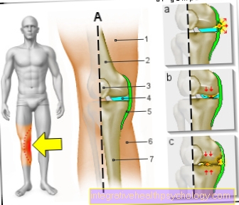
The knee joint is one of the joints with the greatest stress.
Therefore, the treatment of the knee joint (e.g. meniscus tear, cartilage damage, cruciate ligament damage, runner's knee, etc.) requires a lot of experience.
I treat a wide variety of knee diseases in a conservative way.
The aim of any treatment is treatment without surgery.
Which therapy achieves the best results in the long term can only be determined after looking at all of the information (Examination, X-ray, ultrasound, MRI, etc.) be assessed.
You can find me in:
- - your orthopedic surgeon
14
Directly to the online appointment arrangement
Unfortunately, it is currently only possible to make an appointment with private health insurers. I hope for your understanding!
Further information about myself can be found at
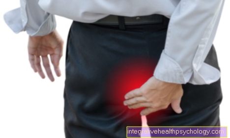
Pain associated with arthrofibrosis
Pain mostly occurs in connection with arthrofibrosis of the knee joint. In most cases, the patient can also assign the pain to the knee joint and, after more specific examinations, specify in which area the pain occurs.
Sometimes, however, pain also radiates. Likewise, by a relieving posture or incorrect load Hip pain occur and one has to look more specifically for the cause in the knee joint and not in the hip.
The pain is often movement-dependent, which means that the pain is more likely to occur when the knee is put under strain, such as when standing or walking. In relaxed postures when sitting or lying down, when the knee is not moved, the pain does not occur or less in comparison.
The pain often responds well to the use of painkillers, so that the sensation of pain can be relieved with appropriate medication.
therapy
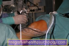
The treatment of secondary arthrofibrosis is surgical. Individual scar strands can easily be removed arthroscopically, which removes the mechanical obstruction. In cruciate ligament surgery, space can be made available for the misplaced graft by expanding the knee roof (emergency plastic surgery) and thereby preventing the graft from hitting again.
The treatment of primary arthrofibrosis is far more difficult and less successful.
In contrast to secondary arthrofibrosis, it often cannot be repaired arthroscopically. In the worst case, multiple arthroscopic operations in particular can lead to a further activation of the chronic inflammatory processes.
For use in symptomatic conservative therapy belong:
- Physiotherapy / physiotherapy
- NSAIDs (non-steroidal anti-rheumatic drugs)
- Physical therapy (heat, cold, Electrotherapy, Ultrasound etc.)
What are the causes of arthrofibrosis?
While for the secondary arthrofibrosis mostly manual operation errors are the cause is the cause of the primary arthrofibrosis still not fully resolved. Different research results are opposed to each other. However, it seems certain that several factors are responsible for triggering and maintaining primary osteoarthritis.
In secondary arthrofibrosis after Cruciate ligament replacement surgery manual operation errors are decisive for a persistent restriction of movement of the Knee joint.
Incorrect placement of the graft can lead to entrapment symptoms (Impingement) of the graft on the roof of the knee joint during knee extension. This problem, which can be observed quite often, is caused by one placed too far ahead Tibia (tibial) Drilling channel. Repeated entrapment during knee extension continuously damages the graft, which can ultimately lead to spherical scarring on the graft (Cyclops Syndrome). The ability to stretch the knee joint is restricted.
In the field of Ankle joints it occasionally comes through a capsule / Torn ligament as part of an ankle trauma (Accident event) to intra-articular (im joint) Scarring in the area of the injured structure or generalized. In this respect, the transition to the secondary Arthrofibrosis to the primary Arthrofibrosis be fluent.
Primary arthrofibrosis is characterized by scarring that involves the entire joint (Connective tissue proliferation).
In addition to this quantitative component, there is the fact that the connective tissue that is formed is also changed in its composition. Connective tissue fibers are properly networked with one another, which further reduces joint mobility.
The following causes excessive scarring are discussed:
Activation and propagation of Fibroblasts (Connective tissue cells) as part of an initial inflammatory process.
- Chronic inflammatory reaction as part of an immunoreactive process.
- Imbalances between pro- and counterflammatory cytokines (Inflammatory messengers).
- Hypoxia - reperfusion injury - theory
(Circulatory disorder) - Genetic factors
To date it has not been clarified which stimuli and in which patients a primary one Arthrofibrosis occurs. Retrospective observations after cruciate ligament surgery could, however, identify risk factors that lead to specific recommendations for the Prophylaxis of arthrofibrosis led.
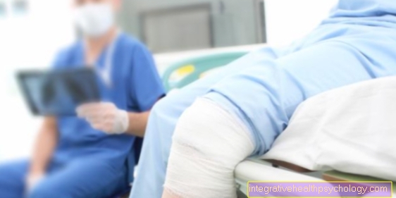
Arthrofibrosis after a knee - TEP
A Arthrofibrosis in the knee joint is a relatively common consequence after surgery on the knee joint (arthroscopic surgery). Such interventions also include Knee - TEP (Total endoprosthesis of the knee joint).
In a knee TEP, the knee joint is through a artificial knee joint replaced. This can result in arthrofibrosis as a consequence of the operation. That means that yourself increased scar tissue forms, which limits the function of the knee joint. A few days to weeks after the operation, the knee joint stiffens, there is increased pain and loading difficulties, or insufficient mobility in the knee joint.
There are different forms of treatment to maintain or improve the mobility of the knee. On the one hand, regular exercise therapy should be carried out as a preventive measure.
Movement and stress on the joint reduce the build-up of scar tissue after the operation. If severe scarring and restricted mobility have already occurred, therapy can be carried out as in other cases of arthrofibrosis (physiotherapy, Anesthesia mobilization, surgical removal of scar tissue).
Differential diagnoses = alternative causes
From the arthrofibrosis other clinical pictures must be distinguished, which also lead to a loss of function of the Knee joint being able to lead.
Rehabilitation deficit (common):
A insufficient postoperative follow-up treatment and too long immobilization (immobilization) can result in a shrinking of the capsule of the knee joint with the result of a persistent restriction of movement. The reasons for this are insufficient postoperative pain elimination, whereby progress in physiotherapy is made more difficult due to pain and insufficient motivation and education of the patient about the importance of postoperative physiotherapy, physical therapy, medical training therapy etc.
Sudeck's disease (rare):
Painful dystrophy (nutritional disorder) and atrophy (shrinkage) of the soft tissues (Musculature, Skin) and bones on the extremities with a typical stage-like course.
The etiology of this disease is still largely unclear.
For more information on this condition, see: Sudeck's disease
MRI of the knee joint
The imaging procedure of choice for the knee joint is this Standard radiograph. The joint and possible changes in the joint space can be assessed. If the cartilage, meniscus, or capsular ligament apparatus are to be rated better, is a MRI (M.agnetrresonancetomography) the method of choice.
This means that an MRI is more of an additional diagnostic option. In the case of arthrofibrosis of the knee joint, it is particularly good that the joints and possible changes can be clearly shown in the MRI and thus a diagnosis can usually be made very reliably.
How can you prevent arthrofibrosis?
Prophylaxis of arthrofibrosis in cruciate ligament surgery:
Due to the difficult therapy If arthrofibrosis has just occurred, the prophylaxis of this disease is of particular importance.
In particular, it was examined which precautionary measures can minimize the risk of developing arthrofibrosis after cruciate ligament replacement.
Prophylactic measures can be taken in preoperative, intraoperative and postoperative Measures are subdivided (modified according to Höher et al. (1999):
preoperative prophylaxis
Choice of the time of operation:
After traumatic Cruciate ligament tear should not be operated on too early. Several studies have shown that one Cruciate ligament replacement surgery within the first 3 weeks after the accident the risk of developing a Arthrofibrosis was significantly increased.
The cause for this is a general "Joint irritation“(Acute traumatic inflammatory reaction) seen through the trauma, with the risk of transition into a chronic one Joint inflammation through additional surgical trauma.
A recovery time of approx. 6 weeks before the operation. At the time of surgery, that should Knee joint freely movable and "irritation-free" (painless, no Joint effusion) be. Accompanying injuries (esp. Internal ligament injuries), should have been treated beforehand. If the knee joint is free from irritation, physiotherapy can be started preoperatively.
Patient education:
The patient must be informed about the severity of the injury and the resulting consequences, especially the postoperative follow-up treatment, and motivated to cooperate.
intraoperative prophylaxis
Operational misplacement of the Cruciate ligament transplant must be avoided at all costs. A common mistake is too far ahead (ventral) placed tibia (tibial) Drilling channel.
Further possible errors are excessively traumatic or long surgery, incorrect placement of the femoral drill channel and incorrect graft fixation.
postoperative prophylaxis
Immediately after the operation physical therapy to be started. An adequate elimination of pain with suitable painkillers is necessary for this. Active and passive (Motor rail) Movement exercises and exercises to mobilize the kneecap are used.
The patient must be motivated to cooperate.
Arthrofibrosis of the shoulder
A Arthrofibrosis can also occur in the shoulder, as the shoulder joint can also be affected here, similar to the knee joint. This leads to pain and restricted mobility. Redness, swelling, or an effusion may also occur. However, these symptoms do not have to occur, the most common being pain and a restriction in possible movements in the shoulder joint.
In the case of arthrofibrosis of the shoulder, the same information applies to diagnosis and treatment as to arthrofibrosis of the knee joint.