AV node
anatomy
Like the sinus node, the AV node is located in the right atrium. However, it lies further down, more precisely at the transition to the right ventricle and thus in Koch’s triangle.
Just like the sinus node, the AV node does not consist of nerve cells, but of specialized heart muscle cells that have the ability to depolarize and thus generate an electrical signal that leads to the heart contraction.
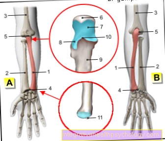
Figure AV node

- AV node
(= Atrial ventricular node)
Nodus atrioventricularis - Sinus node -
Nodus sinuatrialis - Tribe of
Excitation conduction system -
Atrioventricular fasciculus - Right thigh -
Crus dextrum - Left leg -
Crus sinistrum - Rear thigh branch -
R. cruris sinistri posterior - Front thigh branch -
R. cruris sinistri anterior - Purkinje fibers -
Rami subendocardiales - Right atrial -
Atrium dextrum - Right ventricle -
Ventriculus dexter
You can find an overview of all Dr-Gumpert images at: medical illustrations
function
The AV node generated with the help of special ion channels a electrical potential and also receives from Sinus node electrical signals sent, which forwards it to the chamber.
If the sinus node fails, the AV node can step in and the Cardiac contraction continue to take place. Thus, the AV node serves as a filter and in an emergency as generator of electrical potentials. Alone however, he can only do one Heart rate of about 40 strokes generate while the Sinus node a Heart rate from approx. 60 strokes generated.
The Forwarding works through special linesthat pull from the AV node into the ventricle. These lines also consist of specialized heart muscle cells, they pull right and left into the ventricle and end at the apex of the heart. The line from the AV node is called His bundlethat turn into the Tawara thigh split up and into the Purkinje fibers end up.
If the electrical potential now arrives in all heart cells, it can heart contract and Throw out blood. Since the sinus node is connected upstream of the AV node, the atrium contracts shortly before the ventricle and thus helps Filling the chamber with blood. However, 90% of the blood filling takes place via suction that emanates from the chamber.
pathology
It comes to one Disorder the Forwarding of the electrical signals This is called from the AV node to the chamber AV block. There are different degrees of AV block. These are presented under AV block.