Amniotic fluid
introduction
The amniotic fluid is the clear liquid that is in a pregnant woman's amniotic sac, where it helps protect the embryo or fetus.
At an early stage in embryonic development, two separate cavities arise:
The amniotic and chorionic cavities. From the 3rd month, these two cavities merge, the amniotic cavity develops into the amniotic sac and the chorionic cavity into the placenta. Over time, the amniotic cavity steadily increases in volume at the expense of the chorionic cavity. Most of the amniotic fluid in it is taken from the epithelial cells of the amniotic cavity (i.e. fetal tissue) that surround the entire amniotic sac.

Components
The amniotic fluid consists of both maternal as well as from embryonic Shares. The maternal components arrive via the blood through the placenta into the amniotic sac, the embryo releases fluid mainly in the form of urine and also via skin, Lungs and the umbilical cord into the amniotic fluid. Aside from water, the amniotic fluid is made up of various Electrolytes (amongst other things sodium and potassium), Proteins, Lactate, Urea, glucose and also some flaked epithelial cells of the embryo.
Determination of the amniotic fluid
With help of a Ultrasound you can use the amniotic fluid index to determine the amount of amniotic fluid present, which is for everyone Pregnancy check-up should happen.
The normal values are around 30ml in the 10th week of pregnancy, around 400ml in the 20th week of pregnancy and just before birth by 1 liter.
Especially in children who are born late, the amount of amniotic fluid can decrease again towards the end.
The present amniotic fluid is not the same from the beginning to the end of pregnancy. It is subject to a cycle that ensures that the amniotic fluid is completely exchanged within 3 hours, so production and absorption of amniotic fluid should be in balance during a regular pregnancy.
The child drinks from the amniotic fluid, which on the one hand is absorbed through the intestine and reaches the mother's bloodstream through the placenta and on the other hand through the Kidneys is excreted back into the amniotic sac.
Read our article on this: Fruit water examination
tasks
The amniotic fluid fulfills several important functions. On the one hand, because the embryo or fetus practically swims in it, it serves to protect it by absorbing and dampening external impacts to a certain extent. In addition, slight temperature fluctuations can be compensated for by the amniotic fluid. In addition, it enables the unborn to perform movements at an early stage of development and at the same time prevents it from growing together with the cells of the amniotic cavity. Finally, the amniotic fluid also plays a role in the induction of labor as it helps stretch the cervix.
For some years now, amniotic fluid has also been used for another purpose. As part of the Prenatal diagnostics (so one Diagnosiswhich takes place before birth), one can puncture the amniotic sac (Amniocentesis) and remove amniotic fluid. The epithelial cells contained in the amniotic fluid can now be subjected to a chromosome examination. On the one hand, the gender of the child can be determined with a relatively high degree of certainty and, on the other hand, some hereditary diseases and genetic defects, such as the Trisomy 21 (Down syndrome) getting tested. Since this procedure always involves a certain risk, the mother's consent must always be obtained.
Amniotic fluid volume

It is important that the amount of amniotic fluid corresponds to the developmental status of the child and the amniotic sac. If there is too much amniotic fluid in the amniotic cavity, it is called one Polyhydramnios. This can happen, for example, if the fetus has a disability Gastrointestinal passage, does not drink enough, but urine production remains largely constant. If, on the other hand, there is not enough amniotic fluid in the amniotic sac, then there is a Oligohydramnios in front. This condition can be triggered if, for example, insufficient urine is produced due to a malformation in the urogenital tract. The lack of amniotic fluid can ultimately lead to a large number of other malformations, including those of the face, skull, feet or hips, or an underdevelopment of the child's lungs.
In rare cases, an unusually large amount of amniotic fluid passes into the mother's blood during childbirth. This can lead to an amniotic fluid embolism, which is an absolute emergency. The amniotic fluid obstructs small vessels in the mother's lungs, causing shortness of breath and disrupting the coagulation system. As a result, those affected usually have to be ventilated and closely monitored by intensive care.
Color of the amniotic fluid
Amniotic fluid consists of 99% water, along with flaked fetal cells and organic components such as proteins, carbohydrates and fats, as well as electrolytes and urea. The color of the amniotic fluid, like the amount, depends on the week of pregnancy.
At the beginning of pregnancy, the amniotic fluid is usually clear or slightly milky. At birth, the cheese smear turns the amniotic fluid yellowish-cloudy, so-called "Vernix flakes“Become visible. The color of the amniotic fluid thus indicates the child's maturity. In addition, the amniotic fluid can also take on other colors, which can indicate pathological changes. Yellow amniotic fluid is found in cases of blood group incompatibility, whereby the blood group of mother and child do not match. As a result, there is an increased breakdown of red blood cells (Erythrocytes), their breakdown products (Bilirubin) ensure the characteristic yellow color. A flesh-colored coloration of the amniotic fluid is also possible and can indicate the death of the child in the womb. A greenish discoloration of the amniotic fluid is present if the child has their first bowel movement in the uterus (Meconium) has discontinued. This mainly happens when the child is under stress in the womb, as can be the case, for example, when the child is insufficiently supplied with oxygen.
Green amniotic fluid
The Amniotic fluid is produced by the Fruchthöhle itself and is completely renewed approximately every three hours. Usually the amniotic fluid is clear and slightly yellowish colored. Is the amniotic fluid green discolored, this usually indicates that the child was the first in the womb bowel movement has dropped off, who also as a child spook (Meconium) referred to as. This is not uncommon, around 15% of live-born children are born in amniotic fluid containing mekonium. In the majority of cases, however, the first bowel movement is passed in the first few days of life after birth. The cause of the premature stopping of the first bowel movement (meconium) can be one Stressful situation of the child being in the womb, such as Lack of oxygen (Hypoxia). The risk of premature stool in the womb is that amniotic fluid mixed with meconium can get into the child's lungs before or during the birth, which in about 5-10% of cases occurs in a so-called Meconium Respiratory Syndrome can result. Green discolored amniotic fluid mainly indicates one Meconium aspiration if the newborn is flaccid, the skin is bluish instead of rosy, and breathing is greatly reduced. If this is not the case, meconium aspiration is unlikely and can easily be ruled out by the doctor.
Amniotic fluid pH
The amniotic fluid test (A.mniocentesis) is a puncture of the amniotic sac that can usually be performed in women from the 13th week of pregnancy. First, the position of the child is determined with the help of an ultrasound and then a fine needle is passed through the abdominal wall and further through the uterus so that a small amount of amniotic fluid can be removed. Information passes from the child's cells possible hereditary diseases, neurological defects or chromosomal abnormalities like Down syndrome. In addition, parameters such as the PH value, i.e. determine the acidity of the amniotic fluid. Amniotic fluid usually has a pH of 6,5-7, Deviations may indicate a decreased oxygen supply to the child or a bacterial infection.
The pH value of the amniotic fluid differs significantly from that of the much more acidic urine, so that, in case of doubt, the pregnant woman can use rapid tests to determine whether small amounts of urine have leaked, which is not uncommon in the advancing pregnancy, or whether in the frame premature rupture of the amniotic fluid.
Test strips to determine the rupture of the bubbles
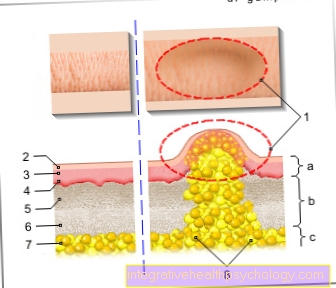
The rupture of the amniotic sac is called the rupture of the amniotic sac, which usually only happens shortly before birth. However, as a result of ascending infections or extreme stress, such as in multiple pregnancies, the amniotic sac can burst several weeks before the calculated due date. A very safe test that the gynecologist conducts to determine the rupture of the bladder is the determination of IGF1, a fetal protein. If the test is positive, the amniotic fluid must have leaked from the amniotic sac and the amniotic sac must have cracked or at least torn. It is important to differentiate between leaked amniotic fluid and leaked urine, as the pelvic floor muscles become increasingly weak in the later months of pregnancy, which can result in slight incontinence.
The difficulty in diagnosis also arises from the fact that the amounts of amniotic fluid are often too small for a reliable diagnosis.
An older test that pregnant women can do at home works with litmus paper. Litmus is a plant pigment that changes color depending on the pH value of the substance applied and thus functions as an acid-base indicator. The litmus paper turns blue with the slightly alkaline amniotic fluid, whereas it reacts red with the weakly acidic vaginal secretions.In addition, there is a large selection of over-the-counter test strips in the pharmacy, also in the form of cotton swabs or gloves, all of which determine the ph value in the vagina and can thus detect premature rupture of the bladder.