Coronary arteries
definition
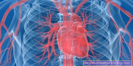
The coronary arteries, also known as the coronary arteries, are vessels that supply the heart with blood. They run in a wreath around the heart and were named after their arrangement.

anatomy
The coronary arteries arise from the main artery, called the aorta, approx. 1-2 cm above the aortic valve. Altogether two branches arise from it, the left and right coronary arteries, these in turn divide into several branches. There are different types of supply, but mostly supplies the right coronary artery, Coronary artery dextra, the back wall of the heart and the partition in the middle of the heart. The Left coronary artery however, supplies the anterior wall, the anterior part of the partition and the outer side of the left ventricle.
Since the left coronary artery divides again after about one centimeter, medicine is often referred to as three coronary arteries. The outgoing branches are called Ramus (Branch) circumflexus and Ramus interventricularis anterior.
Names in Latin
In the case of the arterial coronary vessels, there are two main stems from which other small ones Arteries that supply blood to the heart.
The names of these two arteries are in Latin Left coronary artery, the left, and Coronary artery dextra, the right coronary artery.
The names of the vessels branching off from the left coronary artery are once in Latin the Ramus interventricularis anterior, abbreviated RIVA, and the Ramus circumflexus, RCX for short.
From the right arterial coronary vessel go the Ramus interventricularis posterior, which is abbreviated to RIP, and the Ramus marginalis dexter, also called RMD.
The venous coronary vessels direct the blood into the so-called Coronary sinus. The names of the preceding vessels in Latin are in descending order Vena cardiaca magna, Vena cardiaca media and Vena cardiaca parva.
Supply types
The Ramus circumflexus pulls back and takes care of them left ventricle, during the Ramus interventricularisas the name suggests, between the left and right chambers runs to the apex of the heart and thus the partition and the front part of the heart with it blood provided. The right coronary artery also divides, but there are only smaller branches. The main branch, Ramus interventricularis posterior, pulls back and supplies the rear wall, the Sine- and AV node, the right ventricle, as well as the right atrium plus partly also left ventricular parts.
As mentioned above, there are different types of care. The Legal provider type, the Left-hand supplier type and the "Normal case"which is pronounced at around 70%. Here is a brief summary of the supply for the normal types:
The Left coronary artery provided:
- the left atrium
- the muscles of the left ventricle
- most of the Interventricular septum
- a share of the Anterior wall of the right ventricle
The Coronary artery dextra provided:
- the right atrium
- the right ventricle
- the rear part of the chamber partition
- the sinus node
- the AV node
- a share of the Posterior wall of the left ventricle
function
The Coronary arteries are responsible for the blood supply. The heart is a Hollow muscle, which pumps blood, but is not supplied by it itself. In order to be able to work, it takes, like everyone else muscle, Oxygen and nutrients. This is provided by the coronary vessels, which, thanks to their wreath-like arrangement, supply the entire heart.
pathology
There are a number of them pathological changes (medical: Pathologies) that can affect the coronary arteries.
These include, above all, inflammation, blockages and calcifications.
The Coronary arteries can also "Clog" and trigger different symptoms / diseases depending on the degree of constipation. A Posterior infarction is caused, for example, by blockage of the right coronary artery.
In addition, the unstable / stable Angina pectoris caused by narrowed coronary arteries. The four main risk factors for the so-called Plaque buildup are: Smoke, high cholesterol in blood, diabetes and high blood pressure.
inflammation
Coronary arteries may well be from one inflammation be infested. This can have various causes.
For one, a coronary artery can be part of a Giant cell arteritis, which is primarily the artery affects the temple, but can also manifest itself elsewhere, be inflamed. The special thing about this type of inflammation is the occurrence of Giant cellswhich can be seen in the histological specimen of an affected coronary artery. Besides, she is not bacterial and thus also not purulent.
Also an autoimmune one Polyarteritis nodosa can cause non-bacterial inflammation of the coronary arteries.
In addition, the Leaching of a bacterial germ or that direct introduction of a germ for example through a surgical procedure at heart or through infected foreign material, such as by a Stentcause inflammation in the coronary artery.
Even as part of a arteriosclerosis inflammation of the coronary arteries can occur, for example due to the Appearance of foam cells in the blood vessel.
The symptoms caused by inflammation in the localization are relatively unspecific. That means it can lead to a general feeling of illness Exhaustion and reduced resilience come and also to Pain in the heart. However, these symptoms are usually not that clear and require further detailed information Diagnosis by a heart specialist.
Calcified coronary vessels
If the coronary arteries are calcified, this is usually included in the so-called “arteriosclerosis”. Among other things, calcium is deposited in the blood vessel wall, which consequently leads to hardening of the same.
The cause of the process is usually damage to the innermost layer of the vessel wall.
This can be due to high blood pressure, an immune reaction or a direct wounding of the tissue, for example. In addition, too high levels of bad blood fats, such as triglycerides or LDL (Low densitiy lipoprotein), play a crucial role in the development of a calcified coronary artery, as they promote the inflammatory process and the changes in the blood vessel.
The consequence of a calcified coronary artery can initially be the stiffening and decrease in the width of the blood vessel, and then the innermost layer at this point may tear. The accumulation of coagulant substances at this weak point can further hinder the blood flow and, in the worst case, lead to a heart attack.
- Please also read our detailed article on the subject: Calcification of the coronary arteries
Obstructed coronary arteries
If a coronary artery is blocked, the following heart tissue can are no longer adequately supplied with blood and oxygen and in the worst case it perishes.
This is also known as Heart attack.
A coronary artery can on the one hand slowly getting tighter bit by bit or else it suddenly constipated.
In the first case, the heart muscle supplied by this blood vessel has the possibility of getting blood from so-called blood at an early stage Collateral arteries so that when the main vessel clogs, there is still enough blood and oxygen gets.
However, this is not always the case. One possible cause for this can be Atherosclerosis with a stable plaque be.
In the second case one arrives Blood clot in the coronary artery and clog it acute. Here the muscle tissue of the heart does not really have the opportunity to continue to be supplied with sufficient oxygen and dies. As a result, the performance of the Heart, especially the pumping function, are severely impaired.
Veins
To Care of the heart also belong Veins which are mostly close to the Arteries run away. Your task is to collect the blood again and put it into the right atrium to direct.
The three largest branches are called:
- Vena cardia mediathat with the Ramus ventricularis posterior runs
- Vena cardiaca parvathat runs with the right coronary artery
- Vena cardiaca magnathat with the Ramus ventriculars anterior runs


.jpg)


