Intracranial pressure sign
definition
Signs of intracranial pressure are clinical symptoms and examination findings that indicate the presence of increased intracranial pressure.
These include general symptoms such as headache, nausea and vomiting as well as possibly increased fatigue and loss of appetite. If the increase in intracranial pressure persists over a longer period of time, damage to the optic nerve can result. This causes visual disturbances such as a reduction in visual acuity, which is why these are also considered intracranial pressure signs. The same applies to the detection of a congestive papilla (bulging, reddening and blurring of the optic nerve) when looking at the fundus (ophthalmoscopy).
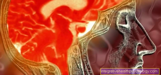
Finally, signs of intracranial pressure, for example in the form of enlarged ventricular spaces (brain compartments that contain the cerebral fluid), can also be made out on radiological images (CT or MRT).

Symptoms
The most important symptoms that can be interpreted as intracranial pressure signs include first
- a headache
- nausea
and - Vomit.
However, these symptoms are very unspecific and, in many cases, can be traced back to a flu-like infection or a gastrointestinal disease.
The additional presence of visual disturbances, which develops over a longer period of time, speaks for an increased intracranial pressure as the cause of the symptoms. Now at the latest it is essential to consult a doctor in order to be able to counteract further consequential damage.
In addition to the symptoms described, many sufferers also complain about
- increased fatigue
- reduced resilience in professional and everyday life
and - increasing loss of appetite.
If an increase in intracranial pressure is not recognized for a long time or is not adequately treated, it can also lead to movement disorders and numbness in various parts of the body. Likewise, increasing hiccups suggests the situation is getting worse.
The additional occurrence of fever and stiff neck suggest the presence of meningitis as the cause of the increased intracranial pressure and should result in immediate intensive care.
Read more on this topic: Signs of meningitis
Hiccups as a symptom of intracranial pressure
Hiccups especially play a role in those affected, in whom a tumor is the cause of the increased intracranial pressure. Even if hiccups may sound harmless at first, they can easily cause significant mental stress for those affected (including sleep disorders). Therefore, a doctor should be consulted if the hiccups are frequent and persistent.
The doctor can initially use possible other symptoms and an eye examination or radiological recordings to estimate how likely the cause is increased intracranial pressure. In addition to the specific treatment of increased intracranial pressure, the hiccups can also be treated separately, for example using proton pump inhibitors (gastric acid blockers such as pantoprazole) or prokinetics (against nausea / vomiting such as domperidone).
Common home remedies (holding your breath, drinking water, etc.) usually only provide short-term relief, if at all.
Also read our articles:
- Hiccups - what to do?
- What are the causes of hiccups?
How do you recognize signs of intracranial pressure on CT?
Since CT images only take a few seconds, they are the method of choice for clarifying a suspected increased intracranial pressure in an emergency, for example as a result of a traumatic brain injury. An expansion of the so-called liquor spaces of the brain is a particularly impressive sign of intracranial pressure in the CT and occurs when the increase in intracranial pressure results from an outflow disorder of the liquor (brain water). The cerebrospinal fluid contained in the liquor spaces is black on the CT, so that the spaces can be recognized in the usual (horizontal) incision in the CT as a butterfly-like structure in the center of the image.
An asymmetry or compression of the liquor spaces speaks for an increased intracranial pressure, then more likely caused by a trauma or a tumor. It is also important to look at the large opening in the skull (foramen magnum). Here, attention is paid to whether the space between the brain stem and the skull bone is normal or reduced, the latter being interpreted as a sign of intracranial pressure. Elapsed cerebral convolutions indicate cerebral edema and are therefore also considered signs of intracranial pressure.
In addition, the cause of increased intracranial pressure can be searched for in the CT: Cause easily recognizable in the CT are, for example, tumors or other obstacles that block the drainage of the cerebral fluid and thus lead to an expansion of the liquor spaces and an increase in intracranial pressure. If such indications are found in the CT, an additional MRI image is often made, which provides more precise images.
Read here how to do that Measure intracranial pressure can
How can you recognize signs of intracranial pressure in the MRI?
Although MRI and CT differ considerably in terms of their functional principle and the representation of different body structures, the same basic rules apply to the determination of intracranial pressure signs in MRI as with CT (see above).
In the MRI image, too, the focus is on viewing the CSF spaces and the space around the brain stem. The MRI usually delivers more precise images and does not involve any exposure in the form of X-rays, but is also significantly more costly and time-consuming than the CT. Therefore, the MRI is sometimes only used if the CT images have not provided a clear finding. In emergencies, in view of the time pressure, CT is preferred anyway.
More on this: MRI of the head
How do you recognize signs of intracranial pressure on the pupil?
Under certain circumstances, evidence of increased intracranial pressure can also be seen when looking at the pupils.
The increase in intracranial pressure can lead to compression of the nerve that is responsible for constricting the pupil (oculomotor nerve). If this nerve is impaired in its function by the compression, a dilated pupil appears on the affected side (or on both sides if the intracranial pressure is increased on both sides). In addition, the pupil reflex, i.e. the narrowing of the pupil in response to the illumination of the eye with an examination lamp, is weakened or even completely failed.
For laypeople, it is sometimes very difficult to assess whether a pupil size and reaction should be considered normal or abnormal. If anything is unclear or if other symptoms such as If the headache or nausea persists for a long time, you should definitely consult a doctor to clarify the matter!
What are chronic intracranial pressure signs?
If the increase in intracranial pressure persists over a longer period of time, chronic damage can occur over time that is difficult or impossible to reverse.
In principle, all kinds of neurological restrictions are possible, from movement disorders (paralysis, coordination disorders) to sensory disorders (numbness, abnormal sensations). However, visual disturbances occur particularly frequently, which are usually expressed in the form of a reduction in visual acuity. In addition, some people affected have impaired color perception and adaptation to dark (adaptation of vision to a dark environment with little incidence of light) may be restricted.
This is how you can recognize signs of intracranial pressure in the baby
Since a baby cannot clearly express its symptoms, the detection of increased intracranial pressure is particularly difficult here.
In general, parents should be alarmed if their infant behaves unusually apathetically or even apathetically (this is typical for an increase in intracranial pressure, but it also occurs with many other serious clinical pictures). Vomiting can also be an indication of increased intracranial pressure, but in the majority of cases it is more likely to be attributed to a gastrointestinal infection. Just like in adults, a dilation of the pupils and a weakening of the light reaction (narrowing of the pupils in response to incident light) are signs of intracranial pressure in babies. In addition, a disproportionately rapid increase in head circumference (according to the rule of thumb, around 1 mm per day is normal) and bulging, bulging fontanelles (cranial sutures) are signs of intracranial pressure that should prompt a medical consultation.
Other possible indications are a stiff neck or an opisthotonus (tilting the head backwards) and difficulty swallowing with frequent belching. Finally, the "sunset phenomenon" should be mentioned: In babies with increased intracranial pressure, a white stripe can often be seen between the upper edge of the iris (the part of the eye that determines the color of the eye) and the upper eyelid.
Read more on the topic: Therapy of the water head
Brain tumor as the cause of increased intracranial pressure
In principle, there is no special intracranial pressure sign that specifically speaks for the presence of a brain tumor as the cause. Nevertheless, there are some observations that are relatively often associated with the cause of a brain tumor.
This includes, for example, a one-sidedness of the described pupil symptoms, i.e. a one-sided dilation of the pupil and an equally one-sided weakened light reaction. Recurring neurological abnormalities (e.g. tingling sensations, movement disorders, numbness) in the same body regions can vaguely steer the suspicion in the direction of brain tumors.
However, only a CT or MRI image can provide final clarity: the images show the brain tumor itself and, often, the asymmetry of the liquor spaces and the displacement of the midline caused by its growth.
Read more on the subject at: Signs of a brain tumor