Right ventricle
definition
The right ventricle is part of the "small" or pulmonary circulation in the right atrium (Atrium dextrum) downstream and pumps the oxygen-poor blood into the pulmonary vessels, where it is again saturated with oxygen in order to then enter the body's circulation via the left heart.

anatomy
The heart is rotated about its longitudinal axis in the left chest cavity, so that the right half of the heart more the anterior chest wall (ventral) is applied while the left half of the heart rather after back (dorsal) shows.
Various anatomical structures can be found inside the chamber:
- The inner surface of the right ventricle is in the area of the Outflow path, i.e. where the blood from the right ventricle reaches the lungs via the pulmonary trunk, smooth-walled,
- of the Rest of the chamber is through protruding muscle bars (Trabeculae carneae) fissured.
In addition, the papillary muscles protrude from the Tricuspid valve into the interior of the ventricle, they are via tendon threads (Chordae tendineae) attached to the valve and prevent it from recoiling into the atrium when the ventricle contracts.
The Wall of the right ventricle is 3-4 mm thick thinner than that of the left ventricle. This is due to the fact that the right heart is essential against you lower pressure, namely the pulmonary pressure prevailing in the lungs of less than 30 mmHg must pump upwhile that left heart when ejecting blood into the aorta against the significantly higher pressure of the body circulationwhich is usually around 120 mmHg, must pump up.
The right ventricle is opened by the ventricular septum (Interventricular septum) separated from the left ventricle, the septum is 5-10 mm thick.
Right ventricle shown

- Right ventricle -
Ventriculus dexter - Right atrial -
Atrium dextrum - Tricuspid valve -
Tricuspid valva - Papillary muscle -
Papillary muscle - Superior vena cava -
Superior vena cava - Lower vena cava -
Inferior vena cava - Pulmonary artery trunk -
Pulmonary trunk - Left pulmonary artery -
Left pulmonary artery - Right pulmonary artery -
Dextra pulmonary artery - Right atrial ventricular valve
(Tricuspid valve) -
Valva atrioventicularis dextra - Left ventricle -
Ventriculus sinister
Small Cardiovascular - (Blue)
You can find an overview of all Dr-Gumpert images at: medical illustrations
function
The heart is functionally divided into a left and a right heart.
The right heart is Part of the "small" cycle (Pulmonary circulation), about the superior and inferior vena cava (Vena cava superior and inferior) the blood gets into the right atrium and from there via the tricuspid valve into the right ventricle.
After contraction of the right ventricle and opening of the pulmonary valve, the blood enters the Pulmonary trunkwho that Blood in the lungs transported where it saturated with oxygen becomes.
The heart action is roughly divided into two sections, the diastole and the Systole.
In the right heart this cycle takes place as follows:
- During the diastole is the Musculature the ventricle relaxed. The AV valve (i.e. the valve between the atrium and ventricle, in the right heart the tricuspid valve) is open and the Chamber is filled with blood.
- The Systole is the phase of Tension. The AV valve is closed, so that with the tension that now follows (contraction) no blood flows from the ventricle back into the atrium.
In the tension phase, the systole, the pulmonary valve is also closed, so the blood remains in the chamber for the time being. Once the Pressure in the chambercaused by the contraction of the muscles, big enough is the pulmonary valve is opened and the Blood flows out of the chamber into the pulmonary circulation.
This heart action, consisting of systole and diastole, runs synchronously in the left ventricle.
Read more on the topic: Task of the heart
Histology wall layering
The Wall layers are set up in the same way in all four interior spaces:
- The innermost layer forms that Endocardium, consisting of a single-layer epithelium, which is lined by the connective tissue lamina propria.
- This is followed by the muscle layer (Myocardium) on.
- The outermost layer forms that Epicardium.
Blood supply
The heart will over the Coronary arteries (Coronary vessels, vasa coronaria) with blood provided.
These are created by the two main vessels, the left and the right Coronary artery (Left and right coronary arteries) and their numerous branches.
This arise from the aorta, directly after their exit from the heart. The right ventricle is mainly supplied by branches of the right coronary artery, but the left coronary artery also provides a small part of the supply.
clinical aspects
The coronary heart disease (CHD) includes clinical pictures caused by a Narrowing of sections of the coronary arteries come about.
The consequence is one Reduced blood flow of the heart. Typical symptom is that Angina pectoris (Herzge).
The maximum variant of the KHK is the Heart attack With complete closure of a section of a coronary artery.
Cause of the CHD are atherosclerotic changes the vessels with Deposits of plaques on the vessel walls. Ruptured (tears) such a plaque and closes the vessel lumen with its contents completely, this leads to heart attack.
A insufficiency of the heart means one Muscle weakness, due to the The heart's pumping capacity is no longer sufficientto provide the body with sufficient oxygen.
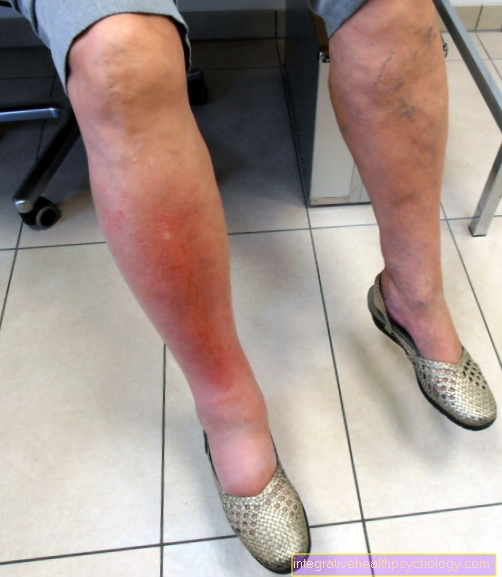
This causes more and more blood to build up in front of the affected section of the heart. At a Right heart failure does this Backwater especially in the form of peripheral edema (Water retention), so edema in the legs is noticeable. Another symptom of heart failure is - among others - a Decline in physical performance.
Causes can be, for example:
- congenital valve defects,
- but also a past heart attack.


.jpg)