Brain aneurysm
definition
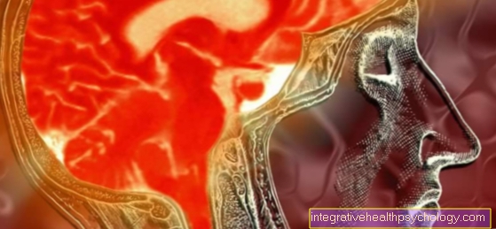
A brain aneurysm is the A bulging blood vessel that supplies blood to parts of the brain. Aneurysms are usually congenital and go unnoticed until they become so large that they press on surrounding tissue or until they tear and cause life-threatening bleeding. They can also develop in the course of life due to increased blood pressure.

The bulging of a blood vessel in the brain is actually not bad, but the danger exists in one at the same time Thinning of the vessel wall. At some point the thinner wall can no longer withstand the pressure of the blood and tears, which too often life-threatening cerebral haemorrhage leading to a stroke. While the majority of strokes are triggered by vascular occlusion, about 10-15% are caused by cerebral haemorrhage. Among the cerebral hemorrhages, the aneurysm is one of the most important causes.
Since brain aneurysms usually do not cause any symptoms before they tear, they go unnoticed for a long time and are often diagnosed as incidental findings.
frequency
Because an aneurysm can go unnoticed for a long time and is often not diagnosed at all, studies on the frequency of occurrence are very vague. In addition, the frequency can also be influenced by factors such as Age and genetic disposition as well as risk factors such as high blood pressure or smoking are influenced.
Studies in patients who were given a vascular imaging of the brain for various reasons showed that around 1-2% of these patients had a brain aneurysm. Studies on autopsied cadavers showed that the frequency is much higher. 7-10% of the examined corpses had an aneurysm in one or more vessels of the brain.
Causes of a cerebral artery aneurysm
There are many known causes of aneurysm formation, and probably many more unknown causes. The basic prerequisite for the formation of this life-threatening bulging of a blood vessel in the brain is this genetic predisposition. Like other connective tissue weaknesses that are inherited, instabilities of the vascular wall can be passed on from generation to generation. However, not everyone with this predisposition ultimately also gets an aneurysm.
Important factors influencing education are Smoking, high blood pressure and also the influence of various hormones. If an aneurysm has formed, it is Blood pressure significantly involved in the further development of the aneurysm. Due to the bulging out of the vessel wall, this is also in place thinner, and can no longer tolerate the pressure that is built up by the bloodstream as well as a vessel wall of normal thickness. If the blood pressure is also increased, the bulge can progress and the vessel wall becomes even thinner. At some point the wall can no longer withstand the pressure and tears down. The so-called Rupture of the aneurysm is a life-threatening emergency as it causes excessive bleeding into the brain.
Signs of a cerebral artery aneurysm
The signs that an aneurysm can trigger in the brain are diverse. The main difficulty is that there is a vascular sac often no complaints or symptoms at all can do if it has not yet ruptured. This is the main reason why aneurysms are often only diagnosed after the hemorrhage or are seen as incidental findings during examinations.
In some cases, however, aneurysms that are not torn cause discomfort and thus prompt an examination. Whether or not an aneurysm causes symptoms depends largely on where in the brain the aneurysm has formed. If the bulge is so large that it exerts pressure on neighboring structures such as other vessels or nerves, this can lead to corresponding complaints. To be mentioned a headache, the frequency and nature of which are new and unusual for the person concerned, some of which are neurological deficits that result from the nerves leading past the sagging blood vessel being stimulated by pressure. Here it can Visual or hearing impairment or impaired movement in various extremitieshow arms or legs come. Depending on the size of the irritated area, it can also Speech disorders come. The neurological abnormalities do not necessarily have to last very long, they can disappear and then reappear a short time later. Also, patients who have an aneurysm in their head can recur for a while dizziness to report. Rarely are all typical symptoms reported at the same time.
Most of the uncracked brain vessel aneurysms do not cause symptoms. The suspicion of an aneurysm is aggravated if one or more Cases in the family of the person concerned is reported. It has been shown that in about 8-10% of aneurysm patients an aneurysm in the family had already become symptomatic.
When a brain aneurysm bursts
The greatest danger of an aneurysm in the brain is not the possible symptoms that can be triggered, but the life-threatening bleeding if the aneurysm ruptures. According to the latest studies, the risk of an aneurysm bursting is not as high as it has been feared over the years.
The risk of an existing aneurysm rupturing depends on a number of factors. The most important factor is the diameter of the aneurysm. Sizes over 7 mm are suspect and tend to burst much more frequently than smaller vascular bulges. An aneurysm would be operated on from a size of 5 mm.
If the aneurysm ruptures, this is an absolute emergency. Because of the high pressure in the arteries, large amounts of blood immediately pour into the space around the ruptured blood vessel. This can sometimes cause serious damage if not treated immediately.
In the case of a torn aneurysm of a cerebral vessel, the most severe headaches are usually reported, which are described as pain of destruction.
Neurological deficits often occur immediately and suddenly. In some cases, people are found unconscious with a ruptured aneurysm. In this case, diagnosis is extremely difficult because the patient cannot provide any information about the symptoms. Immediate notification of an emergency doctor is essential in any case. The patient must be taken to an acute hospital (preferably a hospital with neurosurgery). The vital diagnosis is always a CT of the head. On the one hand, this serves to differentiate between a vascular occlusion and a cerebral haemorrhage, since both can trigger similar symptoms and the therapy is fundamentally different in both cases. The amount of bleeding can also be assessed on the CT.
Read more on this topic: Stroke - how do you recognize it? , What are the consequences of a cerebral hemorrhage
Diagnostics: CT and MRI of the brain in the case of aneurysm

The Immediate action if a ruptured cerebral vascular aneurysm is suspected, it primarily stops CT of the brain The reason is that a CT is faster and provides an overview of whether the clinical picture is bleeding or a vascular occlusion.
A MRI but has the advantage that Vessels better represented and so the extent and spread of the blood can be better assessed. It is therefore at the discretion of the practitioner and the availability of the diagnostic equipment which of the two procedures is used.
If one suspects a vessel has already ruptured, the CT examination is the best diagnostic variant for reasons of time. If one suspects the presence of an aneurysm, an MRI of the brain should be performed for a precise assessment. In magnetic resonance imaging, the patient is pushed into a tube. Before or during the examination, which can take 20-30 minutes, it may be necessary for the person being examined to be injected with a contrast medium. This is necessary when areas in the brain cannot be represented differently. In the case of aneurysm diagnosis, it is always necessary to visualize the vessels with a contrast medium. The contrast medium is injected into the vein via a Braunule and flows into the entire body within a very short time. The blood vessels of the brain are reached within 1-2 seconds. During this time, you should have taken appropriate recordings with the MRT machine in order to make the exact vascular representation possible. The blood vessels are brightly colored, as is a bulge. Existing leaky vessel sections can also be shown through the emergence of contrast medium. In order to be able to plan the operation of an aneurysm carefully, a prior MRI examination of the blood vessels is essential.
Therapy of the aneurysm by coiling
The treatment method for aneurysm called coiling is a relative one new and very effective treatment of a not yet torn aneurysm. If one suspects an aneurysm in the area of a blood vessel in the brain, a vascular imaging must first be carried out. For this purpose a Catheter through the inguinal artery until shortly pushed away from the section where arteries supplying the brain originate. After that, a Contrast media injected and performed an X-ray at the same time. The vessels now appear as if they were in a river bed. Bulges can be clearly recognized by an enrichment of the contrast medium.
If one now decides on a treatment, the coiling process can be used. Here will be platinum-coated, wafer-thin coils are pushed up over the inguinal catheter into the sagging vessel and set down there. The spirals now almost completely fill the sagging area. Within a very short time, the blood flowing past forms small blood clots. The vessel is also said to be thrombosed. Soon the blood clots fill the entire sagging area and seal it off.
The coiling process is very successful. The probability that the vessel will tear open at the coiled point is very low. The coil spirals remain in the vessel forever.
Despite good success, the coiling process also has risks that must be considered and weighed up. When the catheter is advanced, a blood clot can form that is flushed out into the brain, causing a stroke. Another danger of the coiling process is the tearing of the already very thin wall of the sagging part of the blood vessel. This would lead to dramatic bleeding into the brain. Planned thrombosis over weeks can also lead to blood clots being carried over, which can then lead to an acute stroke.
The clipping operation for cerebral artery aneurysm
As an alternative to the coiling process, the so-called OFree clipping operation can be used to treat a cerebral vascular aneurysm. This operation is carried out if the coiling procedure cannot be carried out due to contraindications, or if the aneurysm has exceeded a critical size and can no longer be treated with coiling.
The most common cause of clipping and not coiling is the bleeding of an aneurysm after it has ruptured. Clipping operations can also be planned, i.e. not necessary and with appropriate preparation and planning time.
Advantages and disadvantages in comparison According to the current state of knowledge, clipping and coiling are balanced. The clipping method requires surgery on the open head. To do this, a cut is made behind the hairline and access to the brain is created. Important nerves, such as the optic nerve and important arteries, have to be found and displayed in order to provide an appropriate orientation. As soon as the vessel with the bulge is found, a Clip placed in front of the bulge. The blood is thus prevented from passing through the bagged blood vessel and seeks a bypass circuit. With a dye that is placed in the vessel, the surgeon makes sure that the blood is now looking for another route and that all parts of the brain are adequately supplied with blood.
The operation takes about 3-6 hours depending on the size of the sac and depending on the accessibility and location of the aneurysm. After being monitored in the intensive care unit, the patient is mobilized. A CT scan of the skull is performed on the following day to check for brain swelling after surgery. On the 7th day after the operation, a so-called. Angiology carried out. This is understood to be a vascular representation that is intended to show whether the clip has retained its position and whether the blood flow to the brain is still normal.
Consequences of a cerebral artery aneurysm
In the best case, an aneurysm is discovered and, if it causes symptoms or the risk of rupture is too great, either a coiling or clipping operation can be successfully eliminated without any side effects. Small diameter aneurysms that do not cause symptoms should and can be monitored regularly Last a lifetime without causing problems.
In some cases, however, important neurological structures are either damaged during the procedure, or a ruptured aneurysm causes bleeding into important areas of the brain. The consequences depend on which area was damaged.
The consequences that can occur after a procedure are almost always neurological. They range from Speech disorders, visual disorders, gait disorders, complete paralysis, and coma. It comes to light ones relatively often Unsteadiness and to one Fine motor skills disorder. However, these problems can usually persist within several weeks Rehabilitation measure be remedied in a neurological rehabilitation clinic.
If an aneurysm ruptures, this is a life-threatening emergency. Often, massive hemorrhage causes such severe damage to the brain that the patient permanently loses consciousness. In this case it is fastest possible transport to a hospital with neurosurgery necessary to be able to perform brain imaging and therapy as soon as possible.
If the rupture of an aneurysm survives, 6-8 weeks of follow-up treatment is necessary after the hospital stay. This is mainly through physiotherapeutic and occupational therapy care embossed.
If the patient complains of language problems after aneurysm bleeding or surgery, a large part of the rehabilitation is also done by one Speech therapists designed to ensure that appropriate speaking and language habits are restored as well as possible.
Further consequences of aneurysm bleeding and surgery are follow-up care. Regular follow-up visits by a neurologist or neurosurgeon should be carried out in the first few months to be kept. Furthermore, risk factors should be eliminated or reduced if possible. So should be on the Nicotine consumption should be avoided completely if possible, because the resulting vascular narrowing could make the clip on the vessel unstable. It is also important to have the best possible blood pressure setting; Blood pressure should be checked regularly and, if necessary, blood pressure therapy must be initiated. In addition, any existing diabetes mellitus should be recognized and properly controlled, since poorly controlled diabetes can also affect the blood vessels.
In general, a healthy lifestyle with regular endurance sports, abstinence from nicotine and a Mediterranean diet helps to prevent all types of vascular diseases and their progression, including aneurysm.

.jpg)