Hydrops fetalis
definition
Hydrops fetalis is referred to in prenatal diagnosis as an accumulation of fluid in the fetus. The fluid is located in at least two compartments of the fetus. The edema can spread to large parts of the unborn child's body. The likelihood of hydrops fetalis is 1: 1500 to 1: 4000. Since the suspicion of an accumulation of fluid in the child is an indication of a chromosome change, a malformation of the organs or a serious illness of the fetus, this should be interpreted as a warning sign on the ultrasound.

Causes of fetal hydrops
The most common cause of fetal hydrops is anemia in the unborn child (fetal anemia). This can arise from a rhesus intolerance between mother and child. The rhesus-negative mother produces antibodies against the red blood cells (Erythrocytes) of a rhesus-positive fetus. However, the mother was sensitized earlier, either in a previous pregnancy, through an abortion or a blood transfusion. Ultimately, there is massive damage to the child's erythrocytes and thus anemia in the unborn child. Fetofetal transfusion syndrome and thalassemia are less common immunological causes.
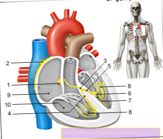
Examples of non-immunological causes that often cause fetal anemia are congenital malformations of the heart. Heart failure can develop as a result of an increase in cardiac output and fluid retention can increase. In addition, infections with toxoplasmosis, syphilis connata, rubella or cytomegalovirus are also among the causes of anemia and thus of hydrops fetalis.
Hydrops fetalis occurs more frequently in chromosomal diseases such as Turner syndrome, trisomy 18 or Down syndrome.
diagnosis
The diagnosis of hydrops fetalis is usually made during a preventive ultrasound examination. You can see the build-up of fluid in the form of a lifting of the skin from the child's body. If the mother has risk factors for developing child anemia, ultrasound examinations should be carried out regularly during the pregnancy. In this way, the pregnancy can be monitored and possibly the development of fetal hydrops can be prevented.
An anemia can also be diagnosed by taking blood from the umbilical cord. If a heart defect is suspected, it can be diagnosed with an ultrasound scan of the heart (Echocardiography) investigate.
Concomitant symptoms
As mentioned earlier, the fetus has a build-up of fluid in the body. Often these are water retention in the abdominal cavity (Ascites) or between the lungs and the chest wall (Pleural effusion).
Another symptom is an increased amount of amniotic fluid (Polyhydramnios). Furthermore, the affected fetus often has a weak heart.
After birth, the children are noticeable for newborn jaundice, anemia and water retention.
Therapy of hydrops fetalis
When treating fetal hydrops, one orientates oneself on the cause. Usually this is caused by fetal anemia, which can be treated in the womb with a blood transfusion through the umbilical cord.
If a fetofetal transfusion syndrome, which causes an uneven distribution of blood between the children, is the cause of the hydrops, the connection in the bloodstream of the twins can be closed again with the help of laser coagulation.
If the cause of hydrops fetalis is a disease with a poor prognosis for the child, it is very important to talk to the treating doctor. He or she can talk to the parents about risks for mother and child as well as the therapeutic options and advise them. Termination of pregnancy may be considered in certain circumstances.
If hydrops fetalis is not treated, this can have consequences not only for the child. The mother can develop maternal hydrops syndrome, which is similar to pregnancy poisoning.
After the birth of a child with hydrops fetalis, this needs intensive medical care. The affected children are often artificially ventilated. They also get blood transfusions and are treated for newborn jaundice by having phototherapy or blood exchange. Any build-up of fluid can be treated with a puncture for relief. Thereafter, the course of therapy depends on the causal disease.
What are the chances of survival and the prospect of a normal life?
Due to the modern diagnosis and therapy options, around 85 percent of children suffering from hydrops fetalis of immunological origin can survive. However, if the cause is non-immunological, fetal mortality can be over 80 percent. In the first trimester of pregnancy, hydrops fetalis often leads to spontaneous abortion. During the third trimester, premature birth, atonic rebleeding, and placental detachment are more common.
In live fetuses, it is very likely to find out the cause of the disease. In rare cases, hydrops fetalis can resolve spontaneously on repeated ultrasound examinations. Also, slight accumulations of fluid can disappear on their own after the birth.
However, in severe cases, it is advisable to terminate the pregnancy as soon as the mother's health is endangered. After birth, artificial respiration is often used so that the affected child can survive. As a rule, the attending physician cannot predict whether the disease will progress positively.