Mucous membrane
Synonym: mucosa, tunica mucosa
English: mucosa
definition
The word "mucous membrane" came directly from Latin "Tunica mucosa" translated. "Tunica" means skin, tissue and "Mucosa" comes from "Mucus" Mucus.
The mucosa is a protective layer that lines the inside of hollow organs such as the lungs or the stomach. It has a slightly different structure than normal skin and has no horny layer and no hair. As the name suggests, this epithelial (= skin) layer is responsible for the production of mucin, or mucin.

Structure of the mucous membrane
The Mucous membrane is as mentioned unkorn, one (e.g. in Intestines) or multilayer (like in the Oral cavity) and can be flat in shape or a elongated, slim basic shape that is taller than it is wide.
Of the three-layer structure is in principle the same in all mucous membranes: the furthest inward one, for cavity showing layer is the Lamina epithelialis mucosae.
She is the real one Epithelial layer. From the outside, the Layer of loose connective tissue and other fibers.
she will Lamina propria mucosae called. It closes on the very outside Lamina muscularis mucosae which made up of a delicate layer smooth muscle cells consists.
To Surface enlargement are so-called Microvilli (finger-shaped protuberances), but also Kinocilia (Cilia) or Stereocilia educated.
The larger the surface, the more the mucous membrane can adhere Nutrients record or exchange this. There are mostly in the mucous membrane Glands, the Mucus (mucilage) and thus keep the tunica mucosa moist.
But there are also mucous membranes, such as the Vaginal mucosa, the glandless is. Here the mucus production is taken over by adjacent sections.
Function of the mucous membrane
The mucous membrane renews itself pretty quickly, about every 3-6 days.
It has a certain barrier function and thus serves to mechanically delimit the organ surface.
Furthermore, the mucosa takes on secretion and resorption processes by transporting molecules into or out of the mucous membrane with the help of active transport proteins.
In addition, the tunica mucosa has lymph follicles, the "mucous membrane-associated lymphatic tissue" or MALT (from English: mucosa associated lymphoid tissue) include.
In this way they can produce certain immunoglobulins, especially a lot of IgA, and protect themselves against pathogens that have invaded.
This defense mechanism should be maintained through a regular supply of micronutrients through food and can be reduced by factors such as stress, environmental pollution (heavy metals, smoking, alcohol, pesticides), medication, too little sleep, etc.
As a result, allergies (hay fever, asthma) as well as bacterial inflammation of the gastric mucosa or bladder infections and also viral diseases of the mucous membranes (rhinitis and bronchitis) can occur.
Chronic inflammation can lead to a thickening of the tunica mucosa, but can also cause other symptoms such as belching, heartburn, diarrhea, bleeding, etc. (for example in the case of chronic gastric and intestinal mucosal inflammation).
Often an operative measure is the result. To avoid this, it is necessary to get the important nutrients through food daily and to avoid bad factors like stress, smoking, bacterial or viral infection, etc. or treat them as soon as possible.
Where is the mucous membrane in our body?
The following mucous membranes can be found in our body: Intestinal mucosa, Uterine lining, Oral mucosa, nasal mucosa, bronchial mucosa, anal mucosa, gastric mucosa and vaginal mucosa.
The oral mucosa
Many internal surfaces of the human body are covered with mucous membranes. A large part of the mucous membrane makes up the surface of the digestive tract. Our food passes several square meters of mucous membrane from the oral cavity to the rectum. The mucous membrane is always structured differently depending on its functional requirements.
In the mouth, the main task of the mucous membrane is to moisten the pulp with saliva and thereby initiate the first step of digestion.
However, only a small part of the saliva is formed by glands in the mucous membrane. The lion's share is formed by the large salivary glands of the head. These include the paired ear, mandibular and sublingual salivary glands.
The mucous membrane of the mouth itself is made up of several layers. A thin layer of cells protrudes into the oral cavity partly keratinized and keratinized squamous epithelium. Horny squamous epithelium is thicker and more resilient than uncornified one. It is therefore found in the areas of the mouth that are exposed to greater mechanical stress from food. An example of this would be the base of the tongue.
The oral mucosa also contains numerous immune cells that protect it from infectious invaders. These include, for example Langerhans giant cellswhich are able to trigger an immune response in the body. With a weakened immune system, for example in the context of an HIV infection or cancer, infections with bacteria or fungi occur more often in the oral cavity. The oral mucosa is then often swollen. So if such an infection occurs, you should always look for the cause of the problem.
Read more on the topic: Swollen lining of the mouth
Next Pigment cells sensory cells can also be distinguished in the oral mucosa. So-called Merkel cells are responsible for the feeling of touch and pressure in the mouth. In this way, the mucous membrane can indirectly pass on the fullness of the mouth to the brain. Other important sensory cells are the taste cells, which are mainly located on the tongue. They enable people to perceive different tastes.
The superficial cells of the oral mucosa sit on a layer of connective tissue that fixes them and holds them in place. In this way, the mucous membrane is not detached when chewing or rubbing the food pulp.
Because the oral mucosa is very well supplied with blood, it can quickly regenerate itself in the event of minor injuries. At the same time, one should make sure that cracks and cuts in the mouth bleed profusely and that they need medical or dental care if necessary.
Gastric mucosa
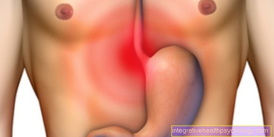
The mucous membrane of the stomach shows some peculiarities that distinguish it from the mucous membranes of the rest of the digestive tract. It is not smooth, but rather raised in longitudinal folds, which smooth out as the stomach becomes full. When viewed greatly enlarged, one can see that the mucous membrane (Tunica mucosa) is not evenly structured. Fields of about 1-5 mm are shown (Gastric area), which lie in a paving stone-like pattern. Small funnel-shaped depressions, called Foveolae gastricae. This is where the gastric glands are located, the roots of which lie deep in the mucous membrane and open into the inside of the stomach. On the one hand, they produce the acidic gastric juice for digestion (see also anatomy of Digestive tract), on the other hand, an alkaline equivalent secretion that protects the stomach from self-digestion. The glandular mucous membrane is only in the main part of the stomach, not at the entrance and exit.
Nasal mucosa
The nasal mucosa consists of the respiratory mucosa (Regio respiratory) and the olfactory mucosa (Regio olfactoria). The respiratory region is named for its function; it represents the first part of the respiratory tract. It covers most of the nasal cavity. They are found on the nasal septum, the side walls and in the turbinates. The uppermost cell layer of this mucous membrane is cylindrically shaped and has kinocilia. Kinocilia are microscopic hairs whose function is to transport dust or secretion towards the throat. Thus they keep the airways free. One of these hairs makes 10 to 20 strokes per second. The respiratory mucosa also contains cells for mucus production and immune defense.
The olfactory mucosa (Regio olfactoria) is found in the upper turbinate, in the nasal dome, and in the upper part of the nasal septum. The primary sensory cells that perceive the smell are located in it. This requires an "olfactory mucus" that is produced by neighboring gland cells (Bowman's glands, Glandulae olfactoriae) is produced. It serves as a kind of detergent that transports odorous substances to the olfactory sensory cells in a soluble form. The mucous membrane of the paranasal sinuses has the same structure as that of the Regio respiratory, but has fewer gland cells.
You might also be interested in: The anatomy of the nose
The lining of the uterus
The uterine lining is also called Endometrium (Tunica mucosa). Lie in her Uterine glands (Uterine glands) that give off an alkaline (basic) secretion. Its function is to protect against infections and to transport the egg cell. Its composition is subject to cyclical fluctuations. The topmost cell layer has a cylindrical structure and has microscopic hairs (cinema cilia and microvilli) that are used to transport the egg cell. The uterine lining is particularly well supplied with blood: it contains spiral arteries, tortuous small blood vessels that change shape depending on the day of the cycle and can increase or decrease the blood supply as required. There are two layers in the uterine lining. The top layer is called Stratum functionalale. It changes over the course of a cycle and is rejected during menstrual bleeding. That lies beneath her Stratum basale. It is not repelled and forms the overlying layer.
Is there a mucous membrane on the eye?
There is no mucous membrane on the eye. What might be colloquially referred to as the mucous membrane is the conjunctiva. It connects the inside of the eyelids with the eyeball and is kept moist by the tear system.
Read more on the subject below: Anatomy of the eye
Mucous membrane of the urethra
The mucous membrane of the urethra is raised in longitudinal folds. From top to bottom it shows three different cell types. The top one is called Urothelium, a layer of cells that is only found in organs of the urinary tract. The middle layer is multi-row and has a highly prismatic shape. The bottom layer is multilayered and uncorned (also found in parts of the oral mucosa, for example). Under the mucous membrane are fine muscle cells that are responsible for continence in the area of the pelvic floor and for the movement of urine in the rest of the urethral area. There are no immune cells or glands in this mucous membrane.
Diseases of the mucous membrane
The mucous membrane plays a role in the following diseases:
- Chronic gastric mucosal inflammation
- Cystitis
- Iron deficiency
- Esophagitis
- Ulcerative colitis
- Crohn's disease
- Celiacia
- Polyps in the nose
- Canker sores in the mouth
- bronchial asthma
- Candidiasis
Inflammation of the mucous membranes
In principle, inflammation can develop on any type of organ or skin and is typically characterized by the following criteria: reddening, overheating, swelling, pain and loss of function. The mechanism behind this is always the same: through damage to the tissues, there is a short-term reduced blood flow and the blood supply is increased by reflex. This leads to swelling and redness. That, in turn, can slow the blood flow and the immune cells Leukocytes (white blood cells) can attach themselves to the scene. They are attracted by certain substances (Cytokines, Interleukins), which mark the damaged tissue as such. This is followed by a variety of repair and / or defense mechanisms in order to restore the function of the organ or tissue.
The best known and most relevant inflammation of the mucous membranes is that of the stomach skin gastritis. It can be acute or (mostly) chronic and have many different causes. The most common is type C gastritis. C stands for chemical and means the long-term use of certain drugs (e.g. aspirin), which destroy the basic mucous membrane protection of the stomach, as the cause. Further classifications are based on A and B; A stands for autoimmunological processes and B for bacterial causes (Helicobacter pylori). An inflammation of the nasal mucous membrane can result, for example, from using a decongestant nasal spray for too long.
Inflammation of the lining of the uterus (Endometritis) is almost always caused by bacteria. The most common pathogens that are known to cause venereal diseases are: chlamydia and gonococci ("gonorrhea"). (Other pathogens are: anaerobes, Gardnerella vaginalis, E. coli, enterobacteria, streptococci, Haemophilus influenzae, mycoplasmas, actinomyces). Mostly it is a question of ascending infections, i.e. diseases of the cervix (Cervicitis), but more rarely diseases descending from the abdomen (such as appendicitis, peritonitis and inflammatory bowel disease). Risk factors for developing uterine lining inflammation are more frequent sexual intercourse with changing partners, low-symptom or untreated genital disorders (Vaginosis or Cervicitis), as well as foreign body implantation (Intrauterine device). At the beginning of menstruation and after childbirth, the protective plug of mucus in the cervix has been lost and therefore also provides an access route for infections. There is also an increased risk of developing endometritis after gynecological or surgical interventions as well as previous pelvic inflammations. The symptoms can vary from mild to life-threatening. The predominant and alarming symptoms here are tenderness, fever and a so-called purulent, creamy discharge.
The inflammation of the urethra is similar to this (see also: Urethritis), as it is often a communicable sexually transmitted disease. The main pathogens are Chlamydia trachomatis and Mycoplasma. The symptoms are again very variable and can be burning, vaginal discharge or creamy-purulent penile discharge in the morning (so-called. Bonjour drops). As with endometritis, the germ should be diagnosed in order to initiate antibiotic therapy. Bacterial inflammation of the oral mucosa is very rare and occurs more in immunosuppressed patients, i.e. patients with a weakened immune reaction. Fungal infestation is more common after antibiotic therapy (Oral thrush; Candidiasis). Chronic inflammatory diseases such as Crohn's disease or venereal diseases such as syphilis can also affect the mouth, but are not among the classic types of infection or key symptoms.
Mucosal erythema
An erythema describes a sharply defined reddening of the skin. It can be found more often on normal skin than on the mucous membrane. There is an infection of the mucous membranes Erythema exudativum multiforme. It is a self-limiting inflammatory reaction that mainly occurs after a viral infection. Self-limiting means that it will heal on its own. It appears mainly on the arms and legs, is target-shaped, burning and itchy. If this is particularly pronounced, the mucous membranes are also affected. Reddening of the mucous membranes in the general sense occurs in many sexually transmitted diseases that are accompanied by inflammation. Also an attack by the fungus Candida albicans (see also: Candidiasis) can include can be described as erythematous (erythema-like).
Mucosal overgrowth
Depending on the function of the individual mucous membrane, it is subject to a more or less pronounced proliferation. It is a so-called unstable alternating tissue. Changes in its shape are therefore mostly wanted by the body.
The term “growth” can mean different growth behavior of cells. Hypertrophy describes the increase in the size of a tissue due to the enlargement of the individual cells. This can affect, for example, the hormonal enlargement of the uterus. Hyperplasia describes a condition in which the number of cells increases and a tissue becomes larger as a result. This affects the hormonal, cyclical build-up and breakdown of the uterine lining (see also: Menstrual period), so it is healthy and wanted (physiological). Its pathological counterpart (pathological) is called Malignancy, so a vicious growth. The term tumor should be differentiated from this. In medical jargon, a tumor describes both swelling due to inflammation or edema, as well as a benign or malignant growth (benign or malignant).
Growths can occur idiopathically (randomly), i.e. without an apparent and disease-related reason. More often, however, they are based on hormonal factors or impaired cell division. In every organ, cell division is limited by intracellular “rules” and barriers (existing within the cell). These mechanisms can be disturbed by prolonged tissue damage. This explains, for example, why years of gastritis (inflammation of the stomach lining) is a risk factor for the development of a malignant ulcer (Carcinogenesis). Sometimes the growth of mucous membrane organs also starts from the glands that are located in the mucous membrane. Then it is the so-called Adenomas, mostly benign tumors.
Growths or swellings due to inflammation are more common and mostly fleeting. For example, a special form of gastric mucosal inflammation (gastritis) the folds of the mucous membrane swell. This disease is therefore also called giant fold gastritis (Ménétrier's disease), it is treated in the same way as a conventional one.
Mucosal cyst
A cyst is an encapsulated, fluid-filled cavity that can in principle arise in any tissue. They can be innate or arise in the course of a lifetime. Congenital cysts are caused by malformation of tissue (for example the dermoid cyst). The other form of cyst, also called acquired cyst, is caused by the blocked drainage of secretions. Since mucous membranes are connected to secretion-forming glands, cysts may develop here. A distinction is made between real cysts (these have their own cell layer as a lining) and false cysts (for example after the tissue has softened due to parasite infestation or other inflammations). If a cyst has been shown to be filled with pus and clearly chambered, it is called an abscess.
The location and formation process of a cyst always play a role in the evaluation of this. Oral cysts, for example, tend to grow progressively, which can then narrow or destroy surrounding structures.A cyst in the bone can dramatically lead to fractures, a mucosal cyst, on the other hand, is less common in principle, as it arises from soft tissue and often becomes symptomatic early on, i.e. causes discomfort. It can be painful if it is caused by inflammation. Congenital mucous membrane cysts in the internal genital tract could reduce fertility through suppressing growth. Can be mistaken for a cyst, canker sores, abscess, erosion, blistering or blistering (vesicle, Bullae) and much more A professional examination by a doctor or dentist is required for a correct diagnosis. Usually, cysts are easy to treat surgically.
Mucosal cancer
Of the types of mucous membrane described, the following cancers are prominent and important: gastric cancer (Gastric cancer), Uterine lining cancer (Endometrial cancer), and cancer of the urinary tract (urothelial carcinoma). Black skin cancer is also found on mucous membranes (Mucosal melanoma) and the mucous membranes of the external genitals can be affected by cancer (vulvar and penile carcinoma; squamous cell carcinoma). As already indicated, diseases of the mucous membranes such as inflammation (gastritis) are important risk factors for the development of cancer in gastric cancer. 90% of them are so-called adenocarcinomas (see also: Colon cancer), which means that the cancer starts from gland cells. Other important risk factors for stomach cancer are alcohol consumption and cigarette smoking, as well as colonization with the germ Helicobacter pylori. At the beginning of the disease, patients usually have few symptoms, rarely unspecific abdominal pain, a feeling of pressure and fullness, and an aversion to meat. This is diagnosed with a gastroscopy including tissue sampling. The only successful treatment is surgery with (in) complete removal of the stomach. Chemotherapy is only given in advanced stages.
Endometrial cancer is the second most common gender-specific cancer in women in Germany. Most women between 60 and 70 are affected. It is now known that the most important risk factor is the long-term intake of estrogens (for example through birth control pills, etc.). This cancer is noticeable early on as painless vaginal bleeding and can easily be diagnosed with a vaginal ultrasound. Affected patients usually have a good chance of recovery. The therapy consists of the surgical removal of the uterus, fallopian tubes and adjacent lymph nodes as well as additional hormonal therapies (progestins).
Urothelial carcinoma is more likely to affect people over 65 and is actually only found in the bladder, the ureter, but rarely or never in the urethra. This cancer manifests itself in blood in the urine, while pain does not go away for a long time. The main risk factor is cigarette smoking. Depending on the stage and location, it can be operated on; in the advanced stage, chemotherapy is used.
A very rare form of black skin cancer affects the mucous membrane. It occurs very rarely because the main risk factor is long-term UV light exposure and the mucous membranes are little exposed to it. It then arises mainly on the uncornified part of the mucous membrane of the lower lip. If a melanoma is detected early, the prognosis is usually excellent with early surgical operation.
Cancer of the mucous membrane of the vulva (external genitalia of women) is a very rare e-disease that affects middle-aged women. It becomes noticeable early on through visual changes, as well as itching, burning and pain, sometimes along with bleeding tears in the mucous membrane. In the early stages, surgery can be used to improve the chances of recovery. As a rule, however, the prognosis is poor and treatment is carried out with radiation or chemotherapy. The counterpart to this in men is, so to speak, penile carcinoma. In both cases, the same cell layer is the exit of the cancer - the squamous cell layer. Penile carcinoma is a very rare cancer that occurs due to poor hygiene and is noticeable early on through hardening or swelling in the area of the glans. A small sample of the skin confirms the suspicion. The only approach to healing is the surgical partial or total excision of the cancer, in later stages also radiation and chemotherapy. Like vulvar cancer, the prognosis is rather poor. Both are associated with human papillomavirus infections (see also: Human papillomavirus), the viruses that also cause cervical cancer and should be vaccinated against girls between 9-13 years old.
Mucosal atrophy
Atrophy is a shrinking of the tissue, either through a decrease in the number of cells or a decrease in the size of the cells. Examples of mucosal atrophies are: Atrophy of the nasal mucosa caused by nasal spray. The decongestant substance xylometazoline removes water from the mucous membrane cells, so there is a brief atrophy. Using the nasal spray for too long (over a week) can permanently damage the cells and cause long-term cell death. The mucous membranes of the female genital tract are subject to hormonal fluctuations in the fertile phases of life. A lack of estrogen in old age, for example, causes atrophy of the vaginal mucosa. Since this is accompanied by the loss of glands and the mucous membranes become drier, they represent a lower protective barrier and the risk of infections increases.
Mucosal folds in the knee
There is no mucous membrane in the knee joint, only several bursa (Synovial bursa). It is a bag-shaped cushion made of synovial fluid, surrounded by thin skin. It lies between muscles and tendons on one side and is bounded on the other by the bone. A bursa can be connected to or separated from the joint cavity. Its function is to improve the sliding of tendons along a bone. Because the knee has so many muscle attachments, there are multiple bursa there. The largest is below that patella (Kneecap) and that Femur (Thigh bone) and is called the bursa suprapatellaris. Other bursa located in the knee are called: Bursa subtendinea musculi gastrocnemii lateralis, Bursa subtendinea musculi gastrocnemii medialis, Bursa musculi semimebranosi, Bursa subpoplitea and many more .. They are each named after the structures that directly surround them.
Mucosal pemphigoid
Pemphigoid is a skin disease in which the upper layer of the skin (epidermis) is lifted from the intact connective tissue underneath due to the formation of bubbles. They are more common on normal skin than on the mucous membrane. Mucosal pemphigoid is a very rare, benign and chronic disease, the origin of which is unclear. Blisters, erosions (superficial tissue defect or tear) and scars form on various skins. Above all, the conjunctiva (then called pemphiguus ocularis) is affected, the further course of which can lead to dehydration and blindness of the eye. It occurs less often in the mouth, on the genitals and in the esophagus. It is to be distinguished from the similar "bullous pemphigoid". Map-shaped redness can be found here (Erythema) with grouped vesicles and bubbles on them. This is an autoimmune disease, i.e. a disease process in which the body's immune system turns against its own structures.
How can you make the mucous membrane swell?
Especially in the winter prepare a swollen lining of the nose Problems. It often occurs with a banal infection of the nasal mucous membrane and is in most cases no risk to health.
Often the swelling goes away with a cold one to two weeks on its own back. However, a swollen nasal lining is commonly called a extremely annoying felt that breathing is impeded during the day and at night. For this reason, we often resort to nasal sprays. These are freely available in the pharmacy and at responsible use harmless to health.
One should at consumption be careful not too much Take nasal spray and also the product to change regularly as the body gets used to the spray and even Dependencies can develop.
Nasal spray often contains something called Zoline. These drugs make the blood vessels in the nasal lining narrow and take care of the decongestant effect. They also work the Against mucus production.
Alternatively to Home remedies be grasped. Popular when there is inflammation of the lining of the nose Salt rinses and inhalation.
Although these bring relief for a short time, they have no effect on the length of the cold. Thus is a balanced use Most likely to recommend sprays and home remedies to reduce the swelling of the mucous membrane.
Mucosal Transplant - What is it?
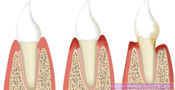
Transplantation is the surgical implantation of foreign or own cells, organs or tissues. If something is removed from one's own body and re-implanted on one's own body, only in another place, one speaks of autologous transplantation (autotransplantation). This is particularly popular with skin transplants. Mucosal transplantation is actually only used in dental or oral surgery treatment (oral surgery is an additional qualification of a dentist and means that he is allowed to operate in the oral area). It is necessary in the event of a mucosal defect, for example after trauma, after the use of implants or after periodontal disease, i.e. after an inflammatory disease of the periodontium (including gum disease, exposed tooth neck). New covering tissue in the form of a transplant may also be necessary after a cancer or a destructive (destructive) infection. Depending on the localization, a sliding flap is possible, i.e. only part of the mucous membrane is cut off and rotated around the tip that remains.
More often, however, a complete flap of the mucous membrane is removed and relocated elsewhere. The mucous membrane of the hard palate is usually used for this because it is coarser in its consistency. So that the new wound produced can heal adequately by itself, a "bandage plate" is put on, a plastic plate that is supposed to protect the open area from irritation, etc. and support wound healing. The free flap can now be sewn on at the required point. Sometimes it is necessary to freshen up the wound edges, i.e. also cut into the actually intact mucous membrane tissue. In this way, blood vessels can grow together from both sides (the place where the flap is inserted and the flap itself) and ensure the blood supply. If the blood supply is insufficient, the flap is rejected. Smokers and diabetics in particular have an increased risk of this. As a rule, however, around 80% of all mucosal flaps / transplants literally heal. The sutures with which the mucous membrane graft is sewn to the desired mucosal site are removed after a week. After 1-2 weeks, the dressing plate can be removed from the palate removal site.


