Medial meniscus lesion
Definition of medial menisal lesion
Under one Medial meniscus lesion one understands one Injury to the medial meniscus. This is located in the knee joint gap and is used for the gliding ability of the Knee joint. There will be a Inner and one outer meniscus distinguished. Both menisci can be injured by accidents or degenerative changes (wear and tear).

Forms of the medial meniscus lesion
It will be different Forms of the medial meniscus lesion distinguished. The medial meniscus can be through a bruise (Contusion) be injured or completely torn (see also: Medial meniscus tear).
Depending on the shape and location of the crack, further shapes are distinguished. A Radial crack refers to a tear that extends from the inner edge along the meniscus radius outwards. If this crack runs parallel to the inner edge after a bend, the radial crack is called Flap tear.
Does the tear line run lengthways through the meniscus, parallel to the main direction of the fibers, this is called a basket handle tear. There is no connection to the inner edge of the meniscus, but the front and rear ends of the fibers connect to the rest of the meniscus.
If the free edge folds into the joint space, this can cause pain. If the crack is horizontal, this is called Horizontal crack designated.
Origin of the cause (etiopathogenesis)

The injury to the inner meniscus can be traumatic (due to an accident) or degenerative (due to wear and tear).
Approx. From the age of 40, degenerative changes occur due to the great stress on the knee joint.
This can lead to spontaneous meniscus tears or to tears due to external force. In contrast, traumatic meniscus lesions particularly affect young people. The cause are usually sports injuries and thus mainly affect young men.
Due to its lower mobility, the inner meniscus is more frequently affected by injuries than the outer meniscus. If, in addition to the medial meniscus, the medial ligament and the anterior cruciate ligament are also ruptured, this is called an "unhappy triad".

I would be happy to advise you!
Who am I?
My name is I am a specialist in orthopedics and the founder of .
Various television programs and print media report regularly about my work. On HR television you can see me every 6 weeks live on "Hallo Hessen".
But now enough is indicated ;-)
The knee joint is one of the joints with the greatest stress.
Therefore, the treatment of the knee joint (e.g. meniscus tear, cartilage damage, cruciate ligament damage, runner's knee, etc.) requires a lot of experience.
I treat a wide variety of knee diseases in a conservative way.
The aim of any treatment is treatment without surgery.
Which therapy achieves the best results in the long term can only be determined after looking at all of the information (Examination, X-ray, ultrasound, MRI, etc.) be assessed.
You can find me in:
- - your orthopedic surgeon
14
Directly to the online appointment arrangement
Unfortunately, it is currently only possible to make an appointment with private health insurers. I hope for your understanding!
Further information about myself can be found at
Symptoms

In the case of an acute meniscus lesion, sudden pain occurs in the joint space, which is particularly pronounced when walking. Possibly is a Snapping over the joint space possible.
If the meniscus flap is trapped in the joint space, this can lead to a Joint blockage to lead. As a rule, the ability to bend and stretch is then eliminated.
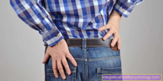
Pain associated with a medial meniscus lesion
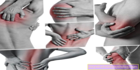
The Medial meniscus lesion typically manifests itself as a sharp pain on the inside of the knee. (see also: internal knee pain)
Pressure on the joint space from above or from the side can make this pain even worse.
Of the Knee pain can arise either directly after an injury or slowly as a result of repeated stress.
In addition, every flexion strain on the knee and turning movements is usually particularly painful. These are also the movements that are most likely to cause a meniscus lesion.
Turning the foot outwards when the knee joint is bent is particularly stressful for the inner meniscus. This movement pattern is also called diagnostic test used when a meniscal lesion appears likely. Lateral pressure on the stretched leg inwards can also increase the pain.
Other movement patterns can also be painful, depending on where exactly the lesion is.
If a meniscus lesion has existed for a long time, the nature of the pain can change significantly:
On the one hand, the pain usually becomes duller here, although more acute pain sensations can occur with exercise. On the other hand, the pain is usually less attributable to a specific side. This is due to the fact that in this case an inflammation and an effusion of the knee joint have often already formed throughout the knee.
If the pain persists for a long time, it often leads to the formation of relieving postures or less strain on the affected knee. This shift in weight can then also overload the other knee and cause damage there. For all symptoms, it is important to have the medial meniscus lesion examined and to determine whether or not surgery is needed.
To treat the pain itself, it can help that knee several times a day too cool and to store high. Taking over-the-counter pain relievers, such as Ibuprofen or Diclofenac on the one hand helps against the pain. Z
to others it also leads to a reduction in the Inflammation in the joint.
In doing so, however, you should make sure that the knees that have been immobilized by the painkillers are not too strained after the pain has ceased. It is also important to take pain medication after an operation. The main concern here is that the pain does not stand in the way of adequate physiotherapy and thus hinder healing. The development of a so-called pain memory should also be prevented in any case. In this respect, if you have knee pain, you shouldn't wait too long to see a doctor and start therapy.
diagnosis
Mostly the anamnesis (medical history) and description of the course of the accident are groundbreaking for the Diagnosis.
On palpation of the joint space one shows Tenderness. In some cases, an accompanying occurs due to the joint inflammation Knee joint effusion on.
There are several Meniscus mark, which when suspected of a Medial meniscus lesion should be checked.
There are several meniscus tests that can result in a positive meniscus sign.
Pain is triggered by passive external rotation of the affected knee joint. This is called positive stone man I sign (positive meniscus sign).
When testing the joint space, the pressure pain that occurs moves backwards (dorsally) when the knee joint is flexed. This is considered a positive Steinmann II sign designated
If the patient sits cross-legged, pain occurs in the joint space. This occurs due to the injured inner meniscus, which exerts pressure on the medial joint space and is called Payr sign designated.
Through the Apley test can also do the Function of the medial meniscus being checked. The patient lies on his stomach while the knee joint is bent by ninety degrees. Pressure on the sole of the foot and external rotation causes pain (meniscus sign) in the medial meniscus lesion.
Further information on this topic can also be found at:
- Meniscus mark
- Meniscus tests
In order to rule out bony injuries, a X-ray made in two levels.
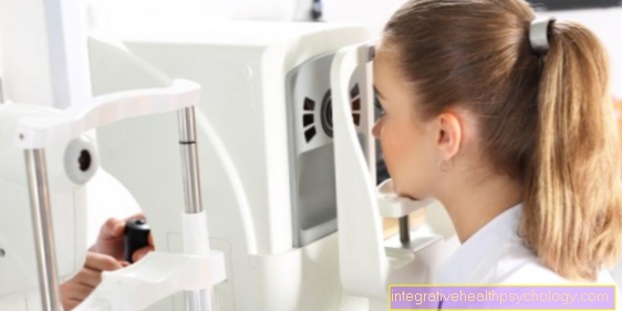
Most valuable in diagnosing a medial meniscal lesion is Magnetic resonance imaging (MRI), which makes it possible to assess the extent of the injury.
Inner meniscus lesion on the posterior horn
The Back horn is the most common place for a meniscus tear in the medial meniscus.
Often a longitudinal crack occurs here, which is usually caused by long-term stress rather than trauma. These longitudinal cracks can in principle heal by themselves. The outer edges of the meniscus are not affected in this form of damage.
However, if the load is passed on, there is a risk that the tear will intensify and thus run through the entire meniscus, or become a so-called Basket handle tear expands.
This can then cause part of the meniscus to fold over and thus lead to a complete blockage of knee mobility.
Horizontal tears also often begin in the posterior horn of the medial meniscus, but these usually occur after an accident. In principle, all of the above-described surgical procedures can be used in this area of the meniscus. A special therapy option that is used primarily for a small lesion of the posterior horn of the medial meniscus is Injection of hyaluronic acid to the affected shares. The hyaluronic acid should help to close the cracks again and the Formation of new cartilage to stimulate.
However, even with this therapy, further exposure harbors the risk of widening the medial meniscus lesion and thus worsening the findings.
Therapy of an inner meniscus lesion
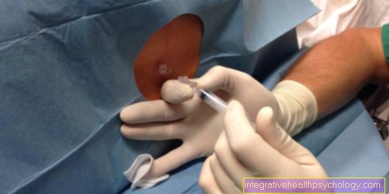
Usually it is part of a Meniscus lesion a Knee joint endoscopy (arthroscopy) carried out. This is not only used for the precise diagnosis of the crack, but also for therapy. Arthroscopy offers different options.
In young patients and cracks in the peripheral third one is tried Meniscus suture perform.
In some cases this is not possible because the medial meniscus tear does not grow together sufficiently. In this case, the meniscus complete or partially removed (menisectomy). If the meniscus has to be partially removed, the procedure should be as gentle as possible, as the meniscus does not grow back. This worsens the shock absorber properties in the knee and one Osteoarthritis of the knee can arise.
In some cases it is necessary to completely remove the meniscus. In this case, the removed meniscus is replaced with a transplant (artificial meniscus). The transplant (artificial meniscus) can consist of an artificial material or be used as part of a direct donation from a corpse. Both materials have advantages and disadvantages, which are still being investigated in studies.
Operation of a medial meniscus lesion
The exact surgical treatment for a lesion of the medial meniscus naturally depends on the exact injury pattern.
Nowadays, however, almost all operations are performed in the form of a knee mirror, i.e. the Arthroscopy carried out.
There are only two small entrances to this procedure Knee joint necessary. This can then be used to repair the damage with the help of instruments introduced.
Usually the damaged parts of the meniscus are then simply removed as part of the arthroscopy. On the one hand, it is important that the largest possible portion of the meniscus remains, on the other hand, not too little should be removed in order to avoid ongoing damage.
Since damage to the medial meniscus is often associated with a Injury to the cruciate ligament or des Inner band These structures may also need to be supplied. Depending on how exactly the damage pattern is on Medial meniscus and depending on the intensity of the stress required after the operation, the tear in the meniscus can also be reattached with the help of a suture.
This is particularly possible when the tear runs close to the meniscus base. Most of them are meanwhile Fixation systems used after the cure do not have to be removed again.
For children in particular, it is advisable to use sutures, as otherwise the risk of further knee damage increases in the long term. However, the follow-up treatment time for meniscus sutures is much longer. A third possibility is the use of meniscus implants. If there is a tear in the peripheral area of the meniscus with no blood supply, a suture is not possible.
However, if the impairment of the meniscus is so severe that it is not possible to simply remove the destroyed parts, an implant may be an option.
This can then take on the support and buffer function that the destroyed meniscus can no longer fulfill. The implants can also usually arthroscopic be introduced. The latest generation of implants usually consists of collagen fibers that are absorbable.
This should then allow the body's own cells to grow in and, in the long term, create meniscus-like tissue. These newly grown tissue can then take over the meniscus function. Overall, the results of the operations are mostly good.
In the case of slight meniscus lesions, conservative therapy is more or less equivalent if the risk is lower.
forecast
The extent of the Meniscus removal, or the meniscus suture determine the prognosis. In the case of a marked removal after a meniscus lesion, one develops quickly Gonarthrosis. This leads to severe discomfort when walking and can be a artificial knee joint (knee prosthesis) make necessary.
As a rule, physical activity must be reduced after any form of meniscus injury.
Time to heal a medial meniscus lesion
The healing time of an inner meniscus lesion differs depending on the surgical procedure.
After a normal arthroscopy, the knee is at least partially loadable immediately after the operation. In this respect, it is also important here to work quickly with a physical therapy and start your own exercises.
To 2-3 weeks can already be started again with light sport, initially most likely by cycling on an exercise bike. Usually after about 6 weeks a full resilience is given again. After a meniscus suture, the healing time is slightly longer. The knee is initially immobilized with a rigid splint for about 1 week.
This is followed by a splint that allows basic movements, but prevents overstretching or excessive flexion in the knee joint. This must be worn for about 6 weeks. Of course, physiotherapy is also important after this operation. However, full resilience is only achieved after about 4-6 months. The healing time and splint treatment with a meniscus implant is roughly comparable to that after a meniscus suture.
Summary
The Medial meniscus lesion is a relatively common injury that occurs particularly in young men as part of athletic injuries. Since the Medial meniscus a less range of motion has as the External meniscus, he is more often affected by injuries.
The meniscus can also tear spontaneously or as a result of incorrect movement in the context of degenerative changes. Depending on the shape of the tear, different types of meniscal lesion are distinguished.
The clinical diagnostics consists mainly in the examination of various clinical signs, which, if they occur, the probability of a Medial meniscus lesion amplify. The diagnosis can also be confirmed by imaging procedures.
If a lesion of the medial meniscus is suspected, a MRI carried out. This serves for an exact diagnosis.
In an arthroscopy (knee examination) the therapy can be carried out in a Meniscus suture or the Removal of the meniscus (Menisectomy).
Depending on the suture or the amount of meniscus removed, a Osteoarthritis of the knee develop.
Figure meniscus tear

Meniscal tear
(= Meniscus rupture)
I - longitudinal tear
II - oblique view (rag tear)
III - radial crack (transverse crack)
IV - basket handle tear (special form)
V - degeneration (wear and tear)
- Inner meniscus -
Meniscus medialis - Outer meniscus -
Lateral meniscus - Posterior cruciate ligament -
Lig. Cruciatum posterius - Anterior cruciate ligament -
Lig. Cruciatum anterius - Femur -
Femur - Kneecap - patella
- Shin - Tibia
You can find an overview of all Dr-Gumpert images at: medical illustrations