Cheekbone fracture
synonym
Zygomatic fracture
definition
A cheekbone fracture is a fracture of the bony cheekbone. The cheekbone is a bone that lies next to and below the eye socket in the area of the upper half of the cheek.
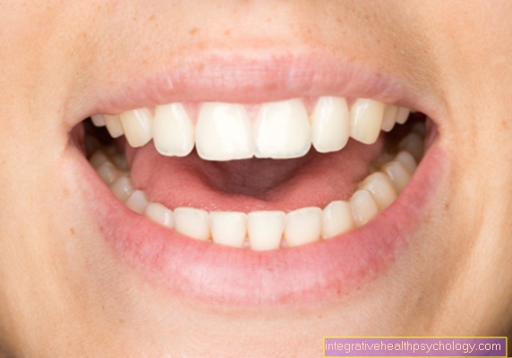
The presence of a cheekbone fracture can often be observed, especially in athletes.
The bony cheekbone is a pair of bones that form the outer boundary of the eye socket. From an anatomical point of view, the cheekbone is one of the so-called facial skull bones.
The bone in the upper part of the cheek can be felt from the outside. In most cases, severe violence leads to a cheekbone fracture. Such a fracture of a facial skull bone can be extremely painful for the affected patient.

causes
The main cause of the occurrence of a cheekbone fracture is a direct, strong, mechanical force on the cheekbone.
Possible mechanisms of an accident could be a collision, a fall or a punch.
The cheekbone fracture is particularly common in soccer players after a collision between two players.
In addition, a cheekbone fracture often occurs in the course of bicycle or traffic accidents. Other possible causes for the occurrence of a cheekbone fracture are physical arguments such as fights.
Depending on the cause and the course of the accident, the cheekbone fracture can take different forms. Above all, the exact location and the resulting bone parts differ greatly depending on the cause. In addition, physical disputes or traffic accidents can impair other bony structures. The zygomatic fracture often occurs in connection with fractures of the nasal bone or the eye socket.
You might also be interested in: Orbital hernia
Symptoms
Since a cheekbone fracture different places of the bone can occur, the Symptoms clearly from each other depending on the exact location distinguish.
Typically, the break line runs from the inner eye socket to the wall of the Maxillary sinus and through the actual zygomatic arch.
In many cases the zygomatic fracture occurs combination With further broken bones in the area of the facial skull.
The typical complaints associated with a cheekbone fracture include Swelling and Bruising in the upper cheek part. In addition, often arise in the course of the cheekbone fracture Hematomas in the Area of the eye (see also: Bruise in the eye). If a cheekbone is fractured, these bruises will only appear in one eye. In this case one speaks of a so-called Monocular hematoma.
Pain in a cheekbone fracture
A cheekbone fracture is described as extremely painful. The cause is usually an extremely strong force on the cheekbone.
The patients feel a strong, intense pain from the moment of impact. In the course of this, it often increases in intensity. Most of the time, the pain is not just limited to the fracture site, but infiltrates into the surrounding area. The patients describe a radiating pain that spreads from the base of the head over the whole face to the lower jawbone. Even slight movements of the facial muscles can trigger extreme pain reactions, so that patients are dependent on effective analgesia.
Due to the radiating pain, it is often not possible to eat properly in the first few days, as even small chewing movements can be painful. The sensation of pain is further supported and intensified by the swellings and bruises that develop, especially in the upper cheek area and around the eyes. This is where hematomas develop, caused by bleeding into the damaged tissue, which are extremely sensitive to pressure.
The violence often causes bleeding, which is also perceived as very unpleasant. On the one hand, the patients bleed from the nose, but it can also bleed into the maxillary sinus. The impairment of vision caused by hematomas on the eyes is also not pleasant for the patient. Usually these are not described as painful, but the occurrence of double images and the blurred vision that can arise, the patients are very limited in the perception of their surroundings. Large hematomas can also be very painful on pressure and restrict the mobility of the eyeball.
It is important to adjust the acute pain well with the help of appropriate analgesia so that further treatment of the fracture, either conservatively or surgically, can be sought.
Patients who have a cheekbone fracture usually describe it strong pain which approximately over the entire affected half of the face can radiate.
It also occurs in many of the affected patients Bleeding from the Maxillary sinus on. This bleeding becomes profuse when it occurs Nosebleed noticeable.
Another typical symptom of a cheekbone fracture is a noticeable one Flattening of the face. The reason for this flattening is usually a change in position (Dislocation) one or more bone fragments. However, this symptom of the cheekbone fracture can easily be overlooked in patients who have severe swelling.
Many of those affected can also have a pronounced asymmetry of Facial skull to be watched. In addition, in the case of a cheekbone fracture, clear steps along the natural course of the bone can often be felt.
Triggered by possible changes in the position of the bone fragments, severe swellings or hematomas, the Eye movement of the patient clearly limited be. In the clinical course, this limitation is mainly caused by the perception of Double vision noticeable.
Since the cheekbone fracture often damages structures in the immediate vicinity of the bone, it can also be damaged Sensory disturbances come. If the maxillary nerve is injured, these sensory disorders are particularly evident in the area of the cheek.
Direct force on the eyes can cause damage in the form of Vision deterioration (blurred vision) to be watched.
How much is the compensation for pain and suffering?
If a cheekbone fracture occurs as a result of an accident that is not their own fault or as a result of violence, for example in the context of a fight, the affected person may receive compensation for pain and suffering. How high this can be cannot be said in general terms, however, but depends on various circumstances, such as the type and severity of the injury and the duration of treatment. Whether and how much compensation for pain and suffering has to be paid is either decided by a court or an agreement can be reached with the other party involved in the accident or the perpetrator. To do this, it is advisable to hire a lawyer. In the event of an intentional injury, compensation for pain and suffering of around 1,000 to 3,000 euros is possible. The amount of a possible compensation for pain and suffering is always determined individually. If a cheekbone fracture occurs as part of a sports injury (footballers or boxers are particularly at risk), compensation for pain and suffering cannot usually be claimed. An exception exists if the person responsible for the violation has committed a serious violation of the rules.
diagnosis

The diagnosis of a cheekbone fracture is usually carried out in several steps. Above all, asking the patient about the exact details Accident mechanism and a detailed doctor-patient discussion (anamnesis) play a decisive role in the diagnosis.
This is followed by a physical examination. In this context, the face of the person concerned is first carefully inspected.
The doctor pays particular attention to this Swelling, bruising and asymmetry in the area of the face.
Then follows a Scanning of the zygomatic arch and the edges of the eye socket. During this part of the physical examination, possible step formation or dislocation of the bone fragments can be felt.
In the further course of the diagnosis of a cheekbone fracture, a radiographic examination of the skull in several planes. If one is suspected concussion or unclear results of the X-ray examination may need an additional Computed Tomography (CT) be performed. In addition, a cheekbone fracture usually follows ophthalmological examination. Depending on the severity of the cheekbone fracture and accompanying injuries, further examinations may be necessary.
Recognize the cheekbone fracture
To recognize a Cheekbone fracture It is important to carefully examine the patient's face during the physical exam.
During the inspection it is noticeable that the affected half of the face is severely swollen. Often times manifest Bruising in the area of the upper cheek, which are often associated with bruises on the eyes.
A hematoma around the eye of the affected side is typical of a unilateral cheekbone fracture (Monocular hematoma). Most patients also experience profuse nosebleeds and bleeding into the maxillary sinus. Often, there are also differences in the symmetry of the two halves of the face. A flattened cheek is also typical of a cheekbone fracture. It is the result of a change in the position of fragments of the bone.
When palpating the bony facial structures, it is typical to feel a step at the edge of the eye socket or at the level of the zygomatic arch. If you suspect a cheekbone fracture, it is important to request an X-ray to determine the extent of the injury and the effects on the surrounding structures. Patients often report that their eyesight has deteriorated.
Typical symptoms are limited mobility of the Eyeball (Globe), blurred vision and Double vision (Diplopia). An ophthalmological examination is therefore always indicated if a cheekbone fracture is suspected. In addition, many patients complain of a sensory disorder in the cheek, as the nerve that supplies the cheek and parts of the upper jaw is often damaged by the high force applied.
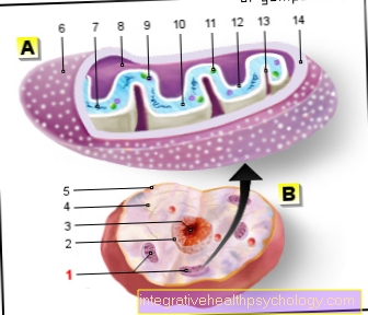
Where is the cheekbone located?
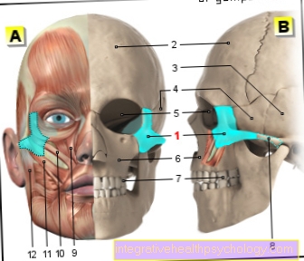
- Zygomatic bone -
Os zygomaticum - Frontal bone - Frontal bone
- Temporal bone - Temporal bone
- Sphenoid bone - Sphenoid bone
- Eye socket - orbit
- Upper jaw - Maxilla
- Molar tooth -
Dens moralis - Zygomatic arch -
Arcus zygomaticus - Upper lip lifter -
Levator muscle
labii superioris - Small zygomatic muscle -
Zygomaticus minor muscle - Zygomatic large muscle -
Zygomaticus major muscle - Masseter (jaw muscle) -
Muscle masseter
A - skull from the front
(Muscles and bones)
B - skull from the left
You can find an overview of all Dr-Gumpert images at: medical illustrations
therapy
The therapy of the zygomatic fracture can either depending on the extent of the injury surgically (conservative) or non-surgical respectively.
Conservative therapy
Patients one not postponed (not dislocated) Cheekbone fractures can be treated conservatively in most cases.
For these patients it is advisable to adhere to a period of several weeks physical protection. In addition, possible swelling in the area of the face can be avoided by careful cooling be treated. In this context, however, it should be noted that the coolant never directly on the skin surface should be applied.
Ideally, the affected patient wraps a flexible one Cooling pad with a kitchen towel and carefully place it on the cheek. In addition, a steady Change between cooling and cold free intervals be respected. This way, the swelling goes down particularly quickly.
Operation of a cheekbone fracture
Surgical treatment is especially necessary if the cheekbone fracture has pieces of bone that have slipped out of their original position.
With the help of special plates and screws, the individual bone fragments can be put back together properly and fixed in their natural position.
The cheekbone fracture operation is usually performed under general anesthesia. In individual cases, however, a surgical procedure can be carried out under local anesthesia.
The typical access to the zygomatic bone is through a small skin incision that extends from the eyebrow to the lower part.
In rare cases, surgical access starting from the oral cavity can also be created.
If the bony eye socket is additionally impaired (especially the outer edge), a skin incision behind the hairline may also be necessary.
During the operation, the slipped bone fragments must first be returned to their natural position.
The individual fragments are then fixed together with the help of small metal plates and special screws.
In patients in whom only the actual zygomatic arch is broken, the natural position can be restored with the help of a special hook technique. In most cases, it is not necessary to insert screws.
Severe injuries involving the eye socket usually require extensive reconstruction. Depending on the exact location of the break lines, additional damage can occur in the area of the vessels. In these cases, aids such as tamponades or balloons often have to be used.
Pronounced defects in the bony structures can also make a transplant necessary. In the case of a cheekbone fracture, pieces of bone or cartilage from the ribs or the hip are often transplanted in addition to foreign materials. The plates and screws used in a cheekbone fracture can be removed after a healing time of about a year. However, another operation must be performed for this purpose.
forecast
A cheekbone fracture can also occur if there are multiple bone fragments and pronounced dislocation in most cases corrected surgically become.
Especially the Aesthetics of the face can be obtained from specialists in oral and maxillofacial surgery at most of those affected fully restored become.
Because of this, the forecast a cheekbone fracture very good. In the majority of patients, a single operation is sufficient to restore the original appearance. In exceptional cases, a second corrective surgical procedure must be performed.
Duration of a cheekbone fracture
A cheekbone fracture mainly affects athletes. For example, in football it can happen that a hard-played ball hits the face of a teammate, who suffers a cheekbone fracture as a result.
The duration of the cheekbone fracture and thus the duration in which the player is absent is often very important for the player personally and for his team.
For the duration of the cheekbone fracture, the player is generally prohibited from participating in training or games. During important games, however, it is possible to protect the face with a face mask and thus to participate in games early after one to two weeks.
If the cheekbone fracture was caused by another sports injury, for example boxing, the patient should refrain from doing sports until the cheekbone has grown stably back together.
Another blow to the zygomatic bone could otherwise break it again more easily. The duration of a zygomatic bone fracture is not only interesting for athletes, but in general for every patient who has a cheekbone fracture (Zygomatic fracture) has suffered.
After the cheekbone has been surgically treated, it takes about 4 weeks for the cheekbone (Os zygomaticum) has grown together and healed.
Although the zygomatic bone is supported by the surgically inserted plate during this time, the zygomatic bone should be spared.
Sports such as boxing or motorcycling, in which the cheekbone is additionally stressed, can prolong the duration of the cheekbone fracture.
In the worst case, the fracture may not heal adequately and the patient may experience permanent mild pain or abnormal sensations (Paresthesia) in the area of the break.
If a patient waits for the duration of the cheekbone fracture until the cheekbone has stably grown back together, the patient can do any sport again after 4 weeks.
Nevertheless, even after the 4 weeks, the patient should make sure that the zygomatic bone is not unnecessarily stressed, as it is after the fracture (the fracture) a fracture in the same area can occur more quickly.
In some patients, there is sufficient stability of the zygomatic bone after 8-9 weeks, which is why endangered sports (Football, handball ...) should be operated with a face mask.
The duration of the cheekbone fracture is therefore individually dependent on the patient. By protecting the cheekbone during the healing phase (for example by lightly chewing), the healing process can be accelerated. This reduces the duration of the cheekbone fracture. However, it can take up to 6 months for the patient to chew and laugh completely symptom-free.
prophylaxis
The Emergence a cheekbone fracture can only occur in the in least cases become.
Special helmets that protect the cheekbone area do not yet exist. Because of this, the prophylaxis (prevention) of a cheekbone fracture as very difficult However, athletes who have recently suffered a cheekbone fracture are advised to wear one special face mask during sporting activity.
How can a mask help?
In the event of a fracture of the cheekbones, wearing a face mask made of carbon can protect the skull from being injured again. Especially in the case of people at risk, such as soccer players, the cheekbones can be protected until the fracture has completely healed. For athletes who want to resume their sport as soon as possible after a cheekbone fracture, a mask can therefore be quite helpful. However, it is essential to consult the attending physician. Only when the edges of the break have healed at least to some extent and the swelling has subsided is it possible to resume training with a carbon mask. However, the mask does not serve the actual healing process. Physical rest is primarily necessary for this. From a medical point of view, it is primarily recommended for both professional and amateur athletes to allow the fracture to heal completely before training or playing is resumed.


.jpg)