Optic nerve
General
The optic nerve (Optic nerve, ancient Greek. “Part of seeing”) is the second cranial nerve and the first part of the visual pathway. It is used to transmit optical stimuli from the retina (retina) to the brain. Because of this, it belongs to the nerves of sensory quality. It runs from the Lamina cribrosa to the junction of the optic nerve, the Optic chiasm, and is about 4.5 cm long.
Development history
The second cranial nerve (optic nerve) as well as the first cranial nerve (bulb and tractus olfactorius) originate from the diencephalon and are thus a protuberance of the brain. Since all other cranial nerves originate from the spinal ganglia of the neural crest, the first two cranial nerves are often referred to as "false cranial nerves".

Emergence
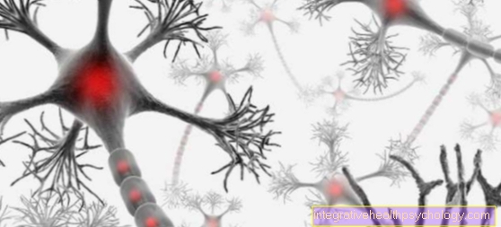
The Axons of the various ganglion cells of the retina unite to form a large nerve, the Optic nerve. For this reason, the optic nerve has no actual core area but three neurons in the retina.
The individual nerve fibers are interconnected with one another. The cells of the Rod and cone layer (1st neuron) be on the Bipolar cells (2nd neuron) and this on the Ganglion cell layer (3rd neuron) interconnected.
The axons of the ganglia then unite to form the large optic nerve (Nervus opticus), which the Retina leaves and takes its course to the brain.
Course of the optic nerve
The course of the optic nerve can be roughly divided into three parts. It starts with one in the eyeball intrabulbar portion, then runs within the eye socket (orbit) (intraorbital part) to finally im skull (intracranial portion) to end.
After the axon union in the Retina the optic nerve leaves the retina at the Optic nerve papilla (Discus nervi optici). Since there are no sensory cells at this point, this point is called blind spot designated. As soon as the nerve exits the retina, it is of the three Meninges and surrounded by the myelin sheaths of the oligodendrocytes. This myelin layer enables the information to be passed on particularly quickly. However, if the optic nerve is damaged, the astrocytes (connective tissue cells) prevent the nerve from regenerating. Of the Optic nerve then continues through the bony eye socket.
It is embedded in fat for protection and enables the Central retinal artery (Central artery of the eye) and the Vena centralis retinae (Central vein of the eye) access to the retina. The two vessels run in the middle of the optic nerve and can enter the retina through the optic nerve papilla. When leaving the eye socket, the optic nerve passes through the Tendon ring (Anulus tendineus communis) of the eye muscles.
After the eye socket, the optic nerve enters the Optic Canal of the sphenoid and is on its way from the Ophthalmic artery accompanied. In the cranial cavity itself, the nerve fibers of the optic nerve run in the subarachnoid space. In front of the pituitary stalk, im Optic chiasm, the nasal nerve fibers of both optic nerves cross. In this way, the signals from the left visual field reach the right hemisphere and vice versa. The partially crossed, partially uncrossed fibers now form the Optic tract. in the Corpus geniculatum laterale are the nerve fibers of the Optic tract switched to the fourth neuron. It then projects its fibers over that Visual radiation (Radiation retinae) the information in the Area striata.
This is the place of the primary vision (primary visual cortex, Area 17). It lies in the area of the back of the head (occipital lobe) and transmits the information to area 18, the secondary visual cortex, as well as to higher visual cerebral cortex areas for further processing.
clinic
Becomes a Optic nerve completely destroyed, the affected eye is blind. However, if only part of the fibers are destroyed, for example in the Optic chiasm, the intersection of the fibers of the right and left eye, the patient suffers from one heteronymous hemianopsia.
This means that the nasal fibers of both eyes fall out, which leads to a restricted field of vision in both eyes on the temporal side (part of the temples). From one contralateral hemianopia one speaks when a Optic tract is affected. The temporal parts of the affected side and the nasal parts of the opposite side are then no longer functional.
Furthermore, the optic nerve can be inflamed (Optic neuritis). Thereby occur an increasing Visual acuity loss (Loss of vision) and possibly a Scotoma (selective visual field loss). Such inflammation is usually caused by demyelinating diseases. Especially those multiple sclerosis can deal with a Optic neuritis manifest.
Due to the inability of the optic nerve to regenerate, restoration of vision is very unlikely.
Diagnosis
The Optic papilla, i.e. the exit point of the optic nerve from the eyeball, can be accessed directly using a Ophthalmoscope to be viewed by an ophthalmologist. Edema in this area indicate severe damage to the nerve and impending blindness.
To differentiate between other diseases at different points of the visual pathway, the Field of view determination (Perimetry). Defects in the visual field, for example nasal deficits, can thus be detected in both eyes and thus damage to the crossed fibers in the Optic chiasm be diagnosed. With help visually evoked potentials (VEP) the nerve conduction velocity of the optic nerve can be determined.
For imaging the nerve and its course, the Ultrasonic (Sonography) that Magnetic resonance imaging (MRI) and that Computed tomogram (CT).
Summary
The optic nerve is that second cranial nerve and, in terms of developmental history, does not belong to the peripheral nerves, like almost all other cranial nerves, but directly to the brain. It is made up of millions of small nerve fibers in the retina and from there it runs to the visual cortex in the brain. On its way through the eye socket, the sphenoid bone and the subarachnoid space into the brain, it is surrounded by a myelin layer and the three meninges. The nasal nerve fibers of both eyes cross in the brain and then continue in the brain as the optic tract. After the passage of the Corpus geniculatum laterale the nerve fibers end in the primary visual cortex (Area 17) at the back of the head (occipital pole).
The further processing of the information then takes place in the secondary visual cortex (Area 18) and the other higher visual cerebral cortex areas. The optic nerve can pass through many places on its way Bleeding, Tumors or other diseases are harmed.
Since the optic nerve is unable to regenerate, recovery of vision is often unlikely. The Diagnosis of optic nerve diseases takes place through the Field of view determination, direct assessment of the optic nerve papilla at the exit point using a Ophthalmoscope or through imaging. The nerve conduction velocity can be determined with the help of the visually evoked potentials measure up.


