MRI of the knee joint
procedure
If a doctor in the hospital or in the practice orders an MRI of the knee, an appointment must first be made. Depending on the order situation and the reason for performing the knee MRI, the affected person may have to wait a few weeks for their appointment.
Before the examination is actually carried out, there is an informative discussion in which the doctor explains the risks of the examination and, above all, has to be informed about previous illnesses and operations.
The actual examination usually only takes about 15 to 30 minutes and is absolutely painless.
Read more on the subject at: Duration of various MRI examinations.

It is important to lie still during the entire duration of the examination so that the images can be used.
The examining doctors have a view of the MRI room during the entire procedure and can talk to and understand the examined patients via an intercom.
The MRI machine itself is very loud during the examination, which is why headphones or earplugs are usually offered to block out the loud noises.
After the images have been created, they have to be evaluated by a radiologist. In most cases, the treating doctor discusses the individual findings with the examined person on site today. However, this is a voluntary service and does not necessarily have to take place
Do I have to be sober for the examination?
In the MRI scan of the knee must be the patient not sober be. The normal intake of food and drink is possible.
In contrast, the patient must appear sober for an MRI examination of the upper / lower abdomen (upper abdominal MRI, hydro MRT).
You can find out more about the topic here: Do you have to be sober for an MRI?
Do I have to take off my knee for an MRI?
An MRI of the knee can on the fully clothed patient respectively.
Only that Removal or storage of all metallic objects (also on clothing) is urgently required. There is a risk that these objects will heat up during the examination or interfere with or distort the MRI images.
Can you do an MRI of both knees at the same time?
An MRI scan of both knees at the same time is available possible in principle, but will usually not carried out. There are several reasons for this.
On the one hand, the radiologist can only bill the health insurance companies for the MRI examination of a knee on the day following the schedule of fees for doctors. On the other hand, an examination of both knees takes a very long time (at least 40 minutes) because the knees have to be examined separately one after the other.
Since the patient should not move during this period and should maintain the lying position, the doctor must decide before the examination whether an examination is possible. Possible criteria for the doctor can be Age, the physical condition and other diseases of the patient.
How far do I have to go from the knee to the tube for an MRI?
Depending on the design, between closed and open MRI machines distinguished.
Usually an MRI scan of the knee places the patient feet first only up to the upper body pushed into the tube. This means that the head is usually outside the tube, so people who are under Claustrophobia Sufferers can also be in a closed MRI tube (with the knee joints).
Transmitter and receiver devices outside the MRT tube are often used to transmit the radio waves and detect the pulses.
In some cases - depending on where the corresponding knee coil is attached in the MRT series, the patient has to put his head into the tube.This should always be clarified in advance, as alternatives must be sought if there is claustrophobia.
The latest MRI tubes have also been built so that they are larger in diameter and therefore no longer appear so restrictive. If necessary, a sedative can be administered by the radiologist. In addition, the patient is always given a push button which he can press in the event of severe discomfort during the examination and thus abort the examination.
Read more about the topic here: MRI for claustrophobia- What are the options?
In any case, it is important that the structure to be examined is identified during the creation of the images is not moved, since the generated images would then not be usable. Blur images, comparable to a photograph.
Do i need contrast media?
Depending on the indication, during the MRI scan of the knee a Contrast media through a vein access injected. In general, with the help of the MRI, a good contrast between the different Soft tissue structures in the knee and thus clearly distinguish them from each other.
To Identification of very fine structures (e.g. smallest cracks in the area of the menisci) or for better representation of perfused areas the administration of contrast medium helps.
Usually stable gadolinium chelates (well tolerated) injected intravenously into a vein in the arm. These accumulate in the knee area, especially in the heavily perfused areas and color them light or white.
In comparison to the areas not supplied with blood (including cartilage tissue), which are stained black, a strong contrast can be set.
Read more about the topic here: MRI with contrast agent- is that dangerous?
Risks of an MRI of the knee
In general, performing an MRI is very safe and usually has no side effects. Risks exist due to the strong magnetic field when certain things are ignored. Because of this, it is important to have one thorough educational talk done by a doctor before the MRI is performed.
Everyone poses a particular problem Metal parts on the body This also applies to things that had to be surgically implanted because of a broken bone, for example. Metallic items that can be removed must be removed before the MRI scan.
If there are metal parts in or on the body that cannot be removed, the MRI examination may not take place and an alternative must be found
Discuss this with the radiologist in advance. Especially when metal implants have been placed on the knee as a result of previous operations, e.g. after a anterior cruciate ligament tear, fracture of the tibial head etc. can be one in many cases MRI of the knee be made. However, it has to be said that no statement can be made about tissue structures directly in the implant, since the image there is erased by the metal implant.
Frequently are thus implanted pacemakers, Bone nails or plates as well Insulin pumps Reasons for not being able to conduct the investigation.
In some MRI examinations, so-called Contrast media used, which in rare cases can lead to allergic reactions. In the case of existing kidney diseases, the treating physicians should also be informed of this before performing an MRI with a contrast agent.
Please also read our topic: Risks with contrast media
indication
In order to better assess certain diseases or to facilitate the diagnosis of unclear knee pain, MRI examinations of the knee are often performed. The MRI findings can also show which therapy is suitable for the individual disease and whether, for example, the indication for an operation is given.
Although bony structures can be assessed less well in the MRI compared to the CT, fractures, tumors or other defects of the bone can also be detected in the MRI.
MRI examinations are ordered particularly often if there is a suspicion that cartilage structures are damaged (cartilage damage).
For example, a damaged meniscus or cruciate ligament tear / collateral ligament tear can be diagnosed well with the help of an MRI. MRI of the knee is also often used with fluids or abscesses in and around the joint.
Ultimately, damage to the ligament and muscular system of the knee joint can also be well recognized with the aid of the MRI.
You might also be interested in this topic: Cruciate ligament overstretched
MRI for a meniscus tear
Probably the most common indication for an MRI is the V.a. for a meniscus tear. (see below)
The meniscus, which acts as a shock absorber between the thigh and lower leg, is subject to natural wear and tear over the course of its life and can tear.
The meniscus tear caused by an accident is much less common.
With an MRI of the knee joint, meniscal tears can be shown relatively reliably. But it is precisely the transition from severe degeneration to degenerative meniscus tear that is sometimes difficult to distinguish radiologically.
MRI for cartilage damage in the knee
To assess the cartilage condition or the cartilage damage in the knee from the outside, the MRI is the best diagnostic option.
Since cartilage has a high proportion of water, it differs significantly from the bone on which it rests.
Higher grade cartilage damage (CM 3 ° and 4 °) can be reliably demonstrated. Smaller cartilage damage and surface roughness are not always clearly visible in the MRI of the knee.
In general, the ability to assess cartilage damage in the knee in an MRI is better when the damage is worse.
Cartilage damage behind the kneecap can be assessed particularly well, since this is where the cartilage in the knee is thickest and the cartilage damage can thus be shown particularly well.
- Cartilage damage in the knee
and - Cartilage damage behind the kneecap
MRI for a V.a. a cruciate ligament tear
The cruciate ligament can be shown very well in the MRI. The anterior and posterior cruciate ligament can be traced over its entire length with appropriate images in the MRI.
In most cases, a complete anterior cruciate ligament tear can also be diagnosed with certainty with an MRI of the knee.
The situation is much more difficult if the cruciate ligament is only torn. Assessing how stable the remaining cruciate ligament is in the MRI remains difficult, even as the image quality improves.
A torn cruciate ligament can lie in an intact tube, especially if the mucous membrane tube is more elastic than the anterior cruciate ligament.
Baker's cyst
At a Baker's cyst is it a Outback of the posterior joint capsulethat collects synovial fluid.
An MRI examination is usually not required to detect the cyst (detection with sonography is possible), but to possible causes of the formation of the cyst to be able to differentiate. Baker's cysts often occur in the context of degenerative meniscus lesions or in the context of chronic polyarthritis.
Read more about the topic here: Baker's cyst in the knee
Contraindication
There are certain contraindications, which is why an MRI examination may not be possible.
As during the investigation no metal parts Any form of may be allowed to be in the room or specifically in the patient non-removable metallic objects is a contraindication for performing an MRI scan in the body.
This includes all types of nails and plates which often remain in the body after bone fracture operations. The same naturally also applies to artificial prostheses such as Hip joint prostheses. Also Pacemaker and insulin pumps may be a contraindication. Further contraindications are Cochlea-Implants, Shrapnel in the body, Neurostimulators, Vascular clips made of metal as well as an existing one in the first three months pregnancy.
Most Knee and hip prostheses, such as Metal implants after a Broken bone usually excludes a knee MRI.
Can I do a knee MRI if I am pregnant?
The effect of radio waves and magnetic field on the embryo is not yet fully elucidated and part of current research. However, all results so far do not indicate any danger to the embryo.
Nonetheless, an MRI scan should be performed in the first trimester of pregnancy if possible avoided become. The need for an MRI scan of the knee during the first trimester of pregnancy should be discussed with the attending physician.
An MRI scan is possible during the rest of the pregnancy. However, if necessary, contrast media should be administered in the smallest possible amounts.
Here you can find more information about: MRI in pregnancy- is it dangerous?
Cost of an MRI of the knee
The costs of performing an MRI scan of the knee are compared to other imaging methods very high. An MRI examination costs depending on the effort about 600-800 € for privately insured persons. In most cases, the examination will be paid for in full by the private health insurance company.
The costs for a MRI of the knee in the legal area is significantly lower and is billed directly with the radiologist with the statutory health insurance (GKV).
As soon as a medical indication for the examination to be carried out, the person concerned does not have to pay for the examination as long as they are insured. This applies to examinations in hospitals as well as to examinations in radiological practices. Overall, MRI examinations take place relatively frequently in Germany in an international comparison.
Only Turkey and the USA carry out more MRI examinations than in this country.
It is repeatedly asked whether the number of examinations carried out is necessary. For this reason, there is increasing demand for stricter indication guidelines, which determine when an MRI examination is to be carried out.
You can find more detailed information on the costs of an MRI of the knee at: MRI costs
Duration of an MRI of the knee
The Duration of an MRI of the knee varies depending on the question and the performance of the device.
In general, the newer the MRT machine and the fewer shifts, the faster the examination is finished.
In general, the MRI examination of the knee can last from 20 - max. 40 minutes go out.
If contrast agent has to be given, the duration can be extended further.
Alternatives to MRI
The investigation of the Knee joint with help of a MRI machine represents in most cases the best possibility is to assess the structures there. There are also structures such as ligaments (especially cruciate ligaments), cartilage (including cartilage damage and meniscus damage) and connective tissue in the MRI of the knee well recognized and differentiated is the investigation with the use of this device in many cases without alternative.
This poses especially for patients with Claustrophobia poses a problem because they have to lie in a small tube for a long time during the examination. This claustrophobia can often be remedied by using new MRT devices that are "open" (open MRT's). Also the Use of sedatives can help reduce these people's anxiety.
The MRI of the knee joint is used in children and Pregnant women preferably carried out because, according to current medical knowledge, the magnetic field has no effects on the human body. In contrast to the use of diagnostic methods which work with X-rays, this is an advantage for the MRI examination of children and pregnant women.
Alternatives to the examination with magnetic resonance tomography must be found if there are contraindications for the examination. In these cases you need imaging tests X-rays such as Ultrasonic can be used.
However, it must be noted that the differentiation of the affected structures when using these examination methods is very difficult or impossible.
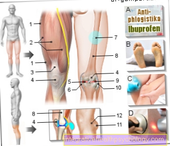
MRI around the meniscus
In the MRI the two face Menisci When viewed from the front, it looks like two wedges, which rest on the right and left of the lower joint part of the knee joint. The view from above shows the meniscus like two C's or crescent moons with the opening facing each other, the outer meniscus is almost closed. In a healthy meniscus, the black of the cartilage is continuous; there should be no bright spots or stripes.
Meniscal tear
An MRI scan is the method of choice when having a Meniscal tear is suspected. If the meniscus ruptures, synovial fluid will flow through the newly created gap, which is bright in the T2 recording technology represents and thus stands out from the surrounding cartilage. Also in the T1 recording technology a light line may be visible in the otherwise dark meniscus, but this can be a sign of degeneration. Furthermore, a changed shape of the surface of the menisci, which is normally smooth and even, can be determined.
Meniscus degeneration
In the case of meniscus degeneration, the MRIthat the otherwise uniform coloration of the cartilage has been removed; light areas, so-called signal increases, appear. The surface of the meniscus is no longer smooth, but rather frayed. In addition, cracks, contour unevenness and detached cartilage parts can be found.
Meniscus damage
At a Meniscus damage the otherwise black structure is no longer continuous, but there are blotchy or streaky areas of light that indicate cracks or damage to the cartilage. The otherwise even surface can be interrupted and frayed.
Degenerative meniscus damage begins centrally and spreads outwards. They can be divided into different degrees:
- Central
- Horizontal, not reaching the surface
- Ribbon-shaped and reaching the surface (from here on one speaks of a meniscus tear)
- Multiple
MRI around the cruciate ligament
Cruciate ligament anatomy
The Cruciate ligaments are best seen in a side view of the knee. They present themselves as thick, arched, dark bands, with the front one Cruciate ligament is narrower than the rear and is displayed a little brighter. The posterior cruciate ligament leads from the articular surface of the thigh bone at the back top down to the articular surface of the tibia. The anterior cruciate ligament leads from the top side to the center of the joint surface.
The cruciate ligament tear
Ligament injuries in the knee Two-thirds affect the cruciate ligaments, with the anterior cruciate ligament being affected up to ten times more often in trauma than the posterior cruciate ligament.
In the MRI image the radiologist can see if a cruciate ligament is torn (here the course of the affected ligament can no longer be traced), whether there is a joint effusion (Fluid build-up in the joint as a result of trauma) or a bruise of the bones and whether there is a possible accompanying injury to the bones.
In addition, injuries to other ligament structures in the knee, especially the menisci, can be assessed.
With a CT or X-ray, however, ligament tears are only visible if they have led to a bone tear. An MRI image can thus complement the clinical examination, whereby it can also lead to an incorrect diagnosis in every fifth case, especially if only a partial tear of a cruciate ligament is present. Therefore, an additional thorough examination by an experienced orthopedic surgeon or trauma surgeon is essential.
Read more on this topic at:
- Cruciate ligament tear
and - MRI for a cruciate ligament tear
General information about MRI

In the Magnetic resonance imaging, short MRI called, it is a radiological, diagnostic method with which the anatomical structures of the body can be shown well. This is an imaging examination that, in contrast to the roentgen or the Computed Tomography (CT) works without the use of harmful radiation.
Some clinical pictures of the Knee joint require an MRI to be performed to diagnose or determine its extent.Since its introduction, the MRT has gained significantly in importance in medical diagnostics. In 2009, almost eight million MRI examinations were performed in Germany, and the number is rising.
Especially "Soft tissue“Like organs or cartilage can be visualized well with the MRI, whereby bones can be visualized better with X-rays.
You can find general information about MRI under our topic: MRI (overview topic)
How an MRT works
Magnetic resonance imaging is based on the Generation of strong magnetic fields and the associated excitation of the atomic nuclei in the body. This enables a very precise representation and differentiation of the types of tissue occurring in the body.
The exact functionality is very complicated and requires an understanding of detailed physical laws.
In contrast to studies that focus on the use of X-rays based, an MRI works completely without the use of harmful radiation.
According to the current state of science and the opinion of most experts, this is the energy that is required to generate the sectional images not harmful to the human body.
There are different examination methods that require the use of an MRT device. With the help of these special examinations, for example, the How the knee joint works examined or the vessels running along the knee are shown precisely. The use of contrast media allows the assessment of the different levels of blood flow different structures and is rarely used in an MRI of the knee.


.jpg)