MRI of the shoulder joint
Procedure of the investigation
The MRI examination usually begins with an informative discussion with the respective specialist. Preparations at home are usually not necessary, but care should be taken to ensure that no fluid or food intake takes place about four hours before the examination. Since contrast media is often used in the shoulder MRI examination for better visualization, the patient is kept sober in the event of a very rare reaction with nausea and vomiting. In the shoulder MRI examination, there is often the exception that contrast agent is given in advance of the actual examination for better assessment. About 10 minutes before the start of the examination, the patient is given the contrast agent either via the vein (indirect arthrography) or in the shoulder joint itself (direct arthrography) injected. The patient is then stopped to move the shoulder joint for 10 minutes.

To partial undressing and the Put down ALL metallic objects from the body, the patient lies in Supine position on a flat examination table, which is then inserted into the MRT machine for approx. 20-minute examination duration is pushed in (the head is also in the device during the shoulder examination). In order to shield the patient from the loud knocking noise during the examination, the patient is usually also given headphone. Furthermore, the patient holds a so-called Emergency button in one hand that can be pressed at any time in the event of problems or complications.
Do I have to be sober during the examination?
With an MRI scan of the shoulder it is not necessary strictly sober (Strictly sobering means no fluid or food intake after 10 p.m. the day before the examination) to appear at the examination appointment. Only from 4 hours before imaging Food and fluid intake should be avoided. The reason for this lies in the possibilitythat during the MRI scan the injection of Contrast media may be necessary. It comes in rarest cases to a Intolerance reaction with nausea and vomitingthen an empty stomach makes things easier. Only in the case of special MRI examinations of the gastrointestinal tract is it necessary to be absolutely or strictly sober before starting the examination.
What clothes can I wear?
As during an MRI examination strong magnetic fields it is important that no metal objects get in or near the examination device.The extent to which the patient is encouraged to take off his or her clothing depends on whether there are metallic objects (buttons, zippers, bra hangers, etc.) on the respective items of clothing. When doing an MRI scan of the shoulder usually a t-shirt or undershirt can be left on, but women in particular are encouraged to use the Take off your bra underneath. It is also important to ensure that Piercings in the area (Nipple piercing, navel piercing etc.) as well away become.
Duration of an MRI scan of the shoulder joint
An MRI of the shoulder joint usually takes about 20 to 30 minutes. The time of preparation is not included here. At a special investigation but it can also 30 to 60 minutes take until the examination is completed. After the examination, the MRI images are printed out or burned on CD and then with the patient discussed.
This also takes about 30 minutes, depending on the extent of the disease. One should look for the full scope of the investigation take about 1 ½ to 2 hours.
The duration of an MRI of the shoulder joint is more of the processes within radiology than dependent on the speed of the MRI machine.
Does an MRI of the shoulder require contrast agent?
Whether contrast media are used always depends on which structures in the shoulder joint are to be examined in the MRI.
There are two ways to administer contrast media. On the one hand, you can put the contrast agent in the vein. However, it is better to administer the contrast agent directly into the joint, which, however, delays the duration of the MRI and is associated with a higher risk of infection.
Contrast media injected especially when one
- Partial tears or tears in the rotator cuff tendons
or - of the long biceps tendons
want to investigate. Also when assessing the bursa over the rotator cuff with regard to bursitis of the shoulder (Subacromial bursitis) contrast agent is required. The contrast agent is injected into the arm through a vein and thus increases the tissue contrast. As a rule, the contrast media are well tolerated. They do not contain iodine and are not chemically similar to the contrast media used in X-ray examinations, which is why they can also be used if you are allergic to X-ray contrast media.
They are excreted in the urine after just a few hours. When examining the cartilage bulge (labrum) Contrast media are also used around the joint socket and articular cartilage.
This contrasts with the examination without contrast agent, which is used for the following structures to be examined:
- with a tumor of the soft tissues,
- with a fracture,
- when the joint changes and
- if you have a joint infection.
Read more on the topic: MRI with contrast agent - is it dangerous?
diagnosis
Magnetic resonance imaging is mainly used when Injuries of the Soft tissues and / or the Tendons be suspected.
Mostly, the MRI of the shoulder joint Tears or Cracks of tendons in the Rotator cuff or the long one Biceps tendon found. Furthermore, you can do a possible Bursitis (Bursitis) recognize or exclude. Does the A tumor is suspected of the shoulder soft tissues, is also used for diagnostics using MRI, as this is X-rays, Ultrasonic (Sonography) or CT (Computed tomography) can't see. The MRI can also be helpful in diagnosing a fracture, a change in the joint or if the joint is infected. In order to make a diagnosis, however, it is always important that, in addition to magnetic resonance imaging, all other examination methods, such as CT, ultrasound and x-rays and a good one anamnese be performed.
Indications for an MRI of the shoulder joint
The indications for an MRI examination of the shoulder include diseases of the muscle-tendon apparatus as well as events in the joint itself and the surrounding soft tissue.
The assessment of fractures (Fractures) in the bones involved in the shoulder joint (upper arm, shoulder blade, collarbone) is also possible, but because of the better representation of bone structures, CT or conventional X-rays are more suitable for this.
One possible indication for an MRI imaging of the shoulder is inflammatory processes in the shoulder joint itself, which can have a wide variety of causes (e.g. infections, autoimmune diseases such as rheumatoid arthritis, metabolic diseases such as gout or activated osteoarthritis in the sense of joint wear).
In the same way, inflammation of the bursa surrounding the shoulder joint (Bursitis) being represented.
Another indication are lesions on the muscle-tendon apparatus of the shoulder joint: this includes diseases of the rotator cuff (e.g. tears in one or more tendons of the four shoulder muscles) and the so-called SLAP lesion (tear in the biceps tendon anchor) glenohumeral instability called (injury to the cartilage lip on the upper edge of the joint socket, where the long biscephalic tendon originates).
Furthermore, a clinically established shoulder instability is a possible indication for an MRI. The instability can be traumatic (e.g. due to an accident), microtraumatic (due to permanent overloading caused by micro-injuries) or atraumatic (e.g. due to a congenital, too wide joint capsule), but also a recurring shoulder joint instability after an already performed operation in this area, may warrant MRI imaging.
In addition, injuries in the acromioclavicular joint (ACG; joint between collarbone and shoulder) indicate an MRI scan. This includes in particular the ACG blast, a lesion in the ligamentous apparatus of this joint.
In addition to these musculoskeletal disorders of the shoulder, the diagnosis of tumorous events in the shoulder area is also viewed as a possible indication for an MRI of the shoulder joint.
Cost of an MRI scan

The Cost of an MRI scan the shoulder distinguish easily between one Private patients and one Cash patients.
The reason for this is that the former are remunerated according to the Fee Ordinance for Doctors (GÖA) and the latter according to the Uniform Assessment Standard (EBM).
The Remuneration rates are lower for statutory health insurance patients than with private patients, which should aim to keep health care costs as low as possible.
Of the Base price for the pure MRI examination of the shoulder joint is at least € 139.89 for private patients and self-payers and at least 124.60 € for statutory health insurance patients.
You can use these basic amounts additional costs be offset, depending on whether additional recordings in special joint positions for special questions or Contrast media is used.
Basically, the costs for an MRI examination of the shoulder joint are only covered by the health insurance companies if there is a reason or an indication for this. Patients who request an MRI examination on their own initiative without a medical need to do so are usually not reimbursed for the costs.
MRI for a tendon tear
Whenever the urgent suspicion of a tendon tear exists in the shoulder (Rotator cuff tear or tear) and the history and physical examination of the shoulder joint suggest it is one special education necessary in order to substantiate or confirm the suspicion and then to be able to initiate optimal therapy.
On the one hand, the Ultrasonic, but especially an MRI image of the shoulder joint.
The latter is very good precisely because magnetic resonance imaging High resolution sectional images generated and especially for the Representation of non-osseous tissue (including tendons) is suitable.
Often there is a during or before the MRI examination Contrast medium administration necessaryto improve imaging and still more accurate representations of the shoulder joint and the surrounding structures.
On an MRI image, the attending physician can then see exactly whether, where and to what extent a tendon is torn, so that a decision can then be made as to whether conservative or surgical therapy is sensible and needs to be initiated.
Anatomy of the shoulder joint
- Definition and structure
The Shoulder joint consists of several parts that articulate with each other (playing together). For one thing, this is the spherical one Humerus head (Caput humeri), which together with the oval articular surface (Glenoid Cavitas) of Scapula (Scapula), the most flexible ball joint in the human body.
This ball joint is so mobile because it hardly a bony lead gives. Since the joint surface of the shoulder blade, due to its oval shape and size, does not fit perfectly with the humerus head, the joint surface must be enlarged. That is why the Cavitas Glenoidalis surrounds one Pan lip 3-4 mm wide (Glenoid labrum).
- The pan lip consists of Fiber cartilage and is attached to the articular surface of the shoulder blade. Furthermore, the Joint capsule of the shoulder joint very slack. This spaciousness and slackness require that downwards (caudal) an approx. 1 cm long Reserve zone (Axillary recess) arises. The recess is only visible when the joint is in a relaxed position.
- Tape apparatus
Of the Tape apparatus the shoulder joint is very weak, although the stress compared to the others Joints is very high. Hence there is no guidance from that Tapes. The joint is held mainly by the strong muscles. Due to the slackness of the ligaments, they occur very often Dislocations (Dislocations), such as muscle- and Tendon tears, due to the heavy use of this.
The following bands (the ligament is called ligament in Latin, it is abbreviated to lig) can be found belonging to the shoulder joint: Reinforcement tape of the Shoulder joint capsule (Coracohumeral ligament). This tape runs from Coracoid process (Bone process of the shoulder blade = scapula) until Humerus (humerus). It is of great importance for the stabilization of the shoulder joint. The Ligament consists of two different parts, one part extending from the humerus to the shoulder blade and the other part from the humerus to another ligament, the Shoulder roof tape (Coracoaacromiale ligament). This ligament is not part of the ligament apparatus of the shoulder joint, but covers the head of the humerus. The second ligament, which is part of the shoulder joint, runs from the socket lip to the joint capsule (Glenoid labrum) (Coracoglenoid ligament) to the bone process of the scapula (processus coracoideus). Third are the Ligg. glenohumeralia. These are several ligaments that run from the edge of the joint capsule to the head of the humerus (Humerus) pull.
- Muscles
All the muscles that support the shoulder joint and make the movements possible are wrapped around the joint like a cuff. That is why we speak of the rotator cuff here when all muscles are summarized. It was made up of four different muscles: The lower bone muscle (infraspinatus muscle), Upper bone muscle (supraspinatus muscle), Subscapular muscle (subscapularis muscle), and the small round muscle (teres minor muscle).
Together with the reinforcement band of the joint capsule of the shoulder joint (Coracohumeral ligament) they form a tough tendon cap that surrounds the joint like a cuff. They run roughly from the shoulder blade (scapula) to the upper arm bone (humerus).
As mentioned at the beginning, the most important task of these muscles is to stabilize the shoulder joint. You push the humerus into the socket. They are also of great importance for various movements: Internal rotation (only with ball or wheel joints. Here the outer side of the arm is turned towards the middle of the body), External rotation (the side of the upper arm facing the body is turned away from the body) and abduction (the lateral spreading of a body part away from the middle of the body). - Bursa
The bursae are very important for the proper functioning of the shoulder joint. Thiefursa subtendinea musculus subscapularis lies under the tendon of the subscapular muscle and ensures that there is no friction between the tendon and the shoulder blade (Scapula) occurs. There is an oval cavity through which the bursa can communicate with the joint capsule. Under the protruding bone of the shoulder blade (Coracoid process) you can also find a bursa that Subcoracoid bursa. It has the function of a reserve space for the joint. The bursa under another protruding bone of the shoulder blade, the acromion (Subacromial bursa) and the bursa under the deltoid muscle (Subdeltoid bursa) together form the subacromial secondary joint. From an anatomical point of view, it is not a real joint, but it supports the abduction of the arm. - Importance of hydrogen in structures for MRI
The human organism consists of around 70% water. Hydrogen is the predominant element in the body. The positively charged water molecules can be magnetized, which the shoulder MRI makes use of. Depending on the weighting (T1, T2, PD), the bone appears black or white on the MRI image because it is particularly low in hydrogen, while the soft tissues are very rich in hydrogen.
In the MRI of the shoulder joint actually only the different proportion of hydrogen atoms is detected and the contrasting and thus the MRT image are produced through differences.
According to their hydrogen content, soft tissues can be very well delimited from one another in MR tomography. They are then displayed in different shades of gray. Malignant and benign tissue can usually be distinguished from one another.

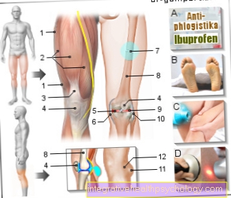
- Humerus head - Caput humeri
- Shoulder joint socket -
Glenoid Cavitas - Shoulder blade - Scapula
- Collarbone - Clavicle
- Shoulder corner - Acromion
- Shoulder-collarbone
Joint -
Articulatio acromioclavicularis - Deltoid - M. deltoideus
- Raven beak process -
Coracoid process - Raven beak extension shoulder corner
Tape -
Coracoacromiale ligament - Joint cavity -
C.avitas articularis - Fiber cartilage ring -
Glenoid labrum - Biceps, long head -
M. biceps brachii - Bursa -
Subacromial bursa - Upper arm shaft -
Corpus humeri
You can find an overview of all Dr-Gumpert images at: medical illustrations


.jpg)