Vestibular nerve
introduction
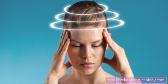
The vestibular nerve is the equilibrium nerve and is part of the vestibulocochlear nerve.
This is the 8th cranial nerve.
The vestibulocochlear nerve can be divided into two parts, the cochlear nerve, i.e. the auditory nerve, and the equilibrium nerve, i.e. the vestibular nerve. The nerve is responsible for transmitting information from the balance organs in the inner ear to the brain.
anatomy
The vestibular nerve originates in the inner ear in the so-called vestibular ganglion.
A ganglion is a collection of nerve cell bodies.
It works its way together with the auditory nerve through the internal auditory canal (Meatus acousticus internus) to get into the posterior fossa. This common course is also referred to as the vestibulocochlear nerve.
The vestibulocochlear nerve enters the posterior cranial fossa at an opening, the so-called porus acousticus internus.
From here the nerve can enter the brain stem at the cerebellopontine angle, where it splits again into the two parts of the vestibulocochlear nerve.
The vestibular nerve then pulls to its cranial nerve nuclei, the "equilibrium nuclei" (Nuclei vestibulares) in the hindbrain (Rhombencephalon). There are a total of four “equilibrium nuclei”, which are named differently depending on their location. There are the nucleus vestibularis superior, the nucleus vestibularis inferior, the nucleus vestibularis medialis and the nucleus vestibularis lateralis.
From here, information that has arrived via the vestibular nerve (so-called afferents) is switched over and forwarded. The information from the organs of equilibrium is passed on to other areas of the brain and spinal cord.
Checking the function of the vestibular nerve
The function of the vestibular nerve can be controlled by means of a Brain stem audiometry, also known as BERA (brainstem evoked response audiometry) must be checked. The test person is exposed to auditory stimuli through headphones in a soundproof room. After the hearing stimuli have been emitted, electrodes attached to the head can normally be used to derive brain potentials, which are then displayed in the form of curves.