Osteochondrosis dissecans therapy
Therapy of osteochondrosis dissecans
The timing of an osteochondrosis dissecans disease cannot be predicted. The different stages of the disease can take place at different speeds. Sudden persistence in a disease stage is possible at any time. Spontaneous healing is also occasionally observed. The rule here is that the younger the patient, the greater the chances of spontaneous healing (especially before the age of 12), but at most approx. 50%.
Overall, the choice of therapy depends on the stage of the osteochondrosis dissecans.
1. Conservative therapy:
Conservative therapy for osteochondrosis dissecans is possible in young patients at an early stage of the disease. A dissection solution must not have taken place yet. Arthroscopically, these areas of the dissection show an intact but softened cartilage covering.
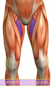
The therapy provides for sports leave and, if necessary, partial relief of the affected leg for 6-16 weeks. MRI follow-ups are necessary to evaluate therapy. Physical therapy measures, physiotherapy, medication, infiltration or nutritional factors have no demonstrable effect on the course of the osteochondrosis dissecans. They are used to treat secondary symptoms (secondary symptoms of osteochondrosis dissecans) such as pain and muscle wasting (muscle atrophy).
Read also the topic: Cartilage flake
2. Operative therapy
Surgical therapy is the method of choice for advanced patients Osteochondrosis dissecans An absolute indication for surgery is when the dissection is dissolved.
The dissection of the dissection represents the maximum damage for the Knee joint On the one hand, the dissecate leaves a hole in the cartilage in its original location; on the other hand, the dissecate as a free joint body damages the still intact knee cartilage. For these reasons, surgical therapy should be used Osteochondrosis dissecans if possible at a stage prior to dissection. The primary goal of all efforts is to maintain an intact cartilage surface.
Operative option 1: The dissecate is not dissolved, the cartilage surface is intact.
- In this case, retrograde or anterograde drillings of the OD area are made with a thin drill (2 mm) performed. The aim is to break through the sclerosis zone and bring about a revitalization of the OD area.With the anterograde bores, from the knee joint side through the intact cartilage thin holes set. The retrograde drilling tries every Cartilage injury to be avoided by drilling into the OD area from the outside. Finding the right place is more difficult here. A postoperative partial load of 6 weeks is necessary.
- Another option is the dead bone to be replaced by the body's own healthy bones. For example, the iliac crest or the tibial head becomes more vital Sponge bones (Cancellous bone) and after hollowing out the dead osteochondrosis dissecans area, it is introduced into this area. This should give the cartilage a vital, stable subsurface again.
Operational option 2: The dissecate is partially or completely detached from the cartilage composite but intact.
- The dissecat is refixed using various fastening systems (screws, pins, bolts). For this purpose, the mouse bed is first freshened so that it can later be waxed in. A postoperative partial load of at least 6 weeks is necessary. After the dissection has grown in, the screws must be removed in a second operation.

I would be happy to advise you!
Who am I?
My name is I am a specialist in orthopedics and the founder of .
Various television programs and print media report regularly about my work. On HR television you can see me every 6 weeks live on "Hallo Hessen".
But now enough is indicated ;-)
The knee joint is one of the joints with the greatest stress.
Therefore, the treatment of the knee joint (e.g. meniscus tear, cartilage damage, cruciate ligament damage, runner's knee, etc.) requires a lot of experience.
I treat a wide variety of knee diseases in a conservative way.
The aim of any treatment is treatment without surgery.
Which therapy achieves the best results in the long term can only be determined after looking at all of the information (Examination, X-ray, ultrasound, MRI, etc.) be assessed.
You can find me in:
- - your orthopedic surgeon
14
Directly to the online appointment arrangement
Unfortunately, it is currently only possible to make an appointment with private health insurers. I hope for your understanding!
Further information about myself can be found at
Operational option 3: The dissecate has come loose, but is no longer suitable for refixation.
- In this case, the therapy can only consist in closing the existing hole in the knee joint cartilage as well as possible. Various methods are available here.
- Pridie drilling / microfracturing
A replacement fiber cartilage tissue is to be stimulated to grow through small holes (see illustration) deep into the healthy bone. In contrast to healthy cartilage, this inferior fiber cartilage grows out of the bone and is supposed to close the hole. - Mosaic plastic / cartilage / bone transplant
Cartilage / bone cylinders are removed from an unloaded part of the knee joint and hammered into the mouse bed using a press-fit technique. - Cartilage cell transplant
In a first step, cartilage cells are removed, cultivated, placed on a carrier medium and, in a second step, transplanted into the knee joint to fill the hole. The procedure is expensive and is not always fully covered by the health insurance company. However, osteochondrosis dissecans in young people is the classic indication for this promising surgical method.