Claustrophobia? - Examination in an open MRI
introduction
In magnetic resonance imaging (in short: MRI) is an imaging process that is used in medical diagnostics primarily to visualize soft tissue and organs. With the help of magnetic resonance tomography, the finest sectional images of the body can be made.
Due to the particularly high-resolution images generated by the MRI, individual changes in organs and soft tissue can be accurately mapped.
General information about MRI can be found under our main topic: Magnetic resonance imaging

The functionality of the MRI is based on a generated inside the device, very strong magnetic field, with the specific Atomic nuclei (especially Hydrogen nuclei / protons) are stimulated in the human body.
The Use of a magnetic field to visualize tissue and organs offers compared to conventional imaging methods enormous benefits.
One advantage of MRI over imaging procedures (for example the roentgen) is the possibility of clearly better soft tissue contrasts to achieve. The differences in the representation of different types of tissue are based on their special fat and water content.
In addition, in contrast to conventional X-rays, MRI images can be without harmful ionizing rays (X-rays) be generated. This has the consequence that it is also with repeated production of MRI recordings not to radiation exposure comes.
Furthermore, there is an improvement in the diagnostic possibilities from the fact that with the magnetic resonance tomograph two different series of pictures can be won. In this context, the Contrast-free- and the Contrast MRI can be distinguished. With the help of the contrast medium MRI, for example inflammatory processes or vital tumor tissue can be better represented by a more intense white coloring.
Next to the ordinary MRI, in which the preparation of the sectional images is a longer period medical diagnostics meanwhile so-called "Real-time MRIs" to disposal. With this form of imaging, individual sectional images in a split second scanned.
With the help of this procedure, for example Movements of organs are displayed true to time. In addition, the real-time MRI during a surgical procedure to represent the exact position of medical instruments be used.
Although magnetic resonance tomography accordingly offers a number of advantages, this form of imaging can, however not in every patient be performed. In patients who have a Pacemaker or implanted defibrillator usually an MRI cannot be performed. The reason for this is the fact that both the pacemaker and the implanted defibrillator during the examination damaged could be. You can also Interactions between these medical devices and the magnetic resonance imaging scanner Significantly harm the health of the patient.
In addition, people who Metal splinters and or Vessel clips made from magnetic materials in an unfavorable position (for example in the area of the Eye or on brain) cannot be diagnosed using an MRI.
Also one Early pregnancy (First trimester; 1.-13. week) is considered a contraindication for performing magnetic resonance imaging. In women who are in early pregnancy, however, must individually decided whether there is any danger to the unborn child.
In addition, performing an MRI scan on people who are under Claustrophobia suffer, be problematic. The reason for this is the fact that with some MRI indications (for example with MRI of the head or MRI of the cervical spine) completely into one closed tube must be laid. With these patients in particular, the enormous advantages offered by MRI have so far only been limited or limited Sedatives be used.
For some time now, however, various radiological institutes have been offering examinations in a so-called open MRI on. Thanks to this new form of imaging, patients with claustrophobia can finally take full advantage of magnetic resonance imaging.
Open MRI
The new open MRI machines are not a tube with an opening at the head and foot end, as has been used in some radiological institutes since the 1990s. Thanks to the new design, which only requires a single support pillar, access to the patient to be examined over 320 degrees is now possible.
The almost unrestricted view from the magnetic resonance tomograph offers enormous advantages, especially for people who suffer from pronounced claustrophobia.
For this reason, an open MRI can be used in almost any patient. In direct comparison to conventional closed MRI devices, however, an open MRI can only generate a significantly lower magnetic field strength. While the magnetic field strength in a tunnel system is around 1.5 to 3 Tesla, only 0.4 to 1.0 Tesla can be generated with open MRI. At first glance, this may appear to be a disadvantage of the open magnetic resonance tomograph. In fact, lowering the magnetic field strength has a major impact on the ability of the device to generate hydrogen atoms (Protons) in the human body. This in turn can have a negative effect on the resolution of the individual sectional images. For this reason, one could assume that the images that were created in an open MRT are much more blurred and less detailed.
According to some studies, however, the reduction in the magnetic field strength can be almost completely compensated for by a longer exposure time. An open MRT is even, if it is a device of more recent year of construction, by increasing the measurement times able to generate equivalent or even better quality cross-sectional images.
In addition, eccentrically located body sections, for example the shoulder, elbow and wrist, can be examined in much more comfortable positions due to the various storage options. In this way, movement artifacts that negatively affect the image quality can be avoided. This is not easily possible in most of the conventional magnetic resonance tomographs (MRT).
Read about this too MRI - How far do I have to put my head in?
Advantages of the open MRI

A open MRT offers a multitude of advantages compared to conventional closed MRI tubes.
To the main advantages This novel diagnostic procedure includes the fact that the open MRI:
- For all examinations suitable is
- performing a comfortable examination guaranteed
- for patients with Claustrophobia suitable is
- For children suitable is
- For elderly people suitable is
- diagnostic examinations themselves severely obese patients can be carried out
- a high image quality supplies.
Although an open MRI is only one compared to a closed tube lower magnetic field strength can generate can through the Adjusting the recording time all examinations are carried out.
An open MRI can Sectional images of the joints, of the internal organs, of the annoy and Vessels and the female breast in particularly high image quality produce. Without radiation exposure, like for example at conventional radiographs or the Computed Tomography acts on the patient, the inner tissue of the human body can be represented and The smallest changes are particularly visible be made.
In addition, an open MRI, in comparison to a closed magnetic resonance tomograph, has a much wider lying surface. This is why the open MRI caters to the patient significantly more comfort. Especially for people who are under pronounced claustrophobia (claustrophobia), the examinations can be carried out without fear attacks thanks to the 360-degree all-round view. Taking one Sedative in these patients is usually prior to examination unnecessary. For this reason, it is no longer necessary to bring an accompanying person to the MRI appointment.
Also for them Examination of Young Children an open MRI provides enormous advantages. If it is not possible to position the child alone in the device, a Parent on the examination couch go. That way you can even with small children high-resolution sectional images can be generated without movement artifacts. The examination in the open MRI is much more relaxed and less stressful for the child.
Due to the extensive design of an open MRT with a 360-degree view, there are also older patients are much more comfortable in the examination device. This benefit can also be used for those with restricted range of motion be used.
Very obese patient could only be examined to a limited extent or not at all in conventional magnetic resonance tomographs. Due to the expansion of the space on the bed surface, these patients can now also be examined with an open MRI without any problems.
In addition, the quality of the cross-sectional images can be reduced by the ability to see the patient to be examined on the Move the examination table in all directions to be able to be positively influenced. The reason for this is the fact that the resolution of every magnetic resonance tomograph central highest is. So, for example, should the liver are displayed, the patient to be examined can be positioned so that the liver is in the center of the MRT.
Disadvantages of the open MRI
Even with techniques that are getting better and better, the lower field strength of the magnetic field cannot compensate for the decrease in quality compared to closed MRI.
Cost of an open MRI
The costs for an open MRI examination not covered by the insurance carriers in all cases (especially in statutory health insurance).
Patients who privately insured are, they can total costthat are incurred when performing an open MRI examination, have the insurance settled.
The statutory health insurance companies reimburse the cost on the other hand only in selected cases. The prerequisite for assuming the costs is what is known as the open MRI Declaration of assumption of costs.
This is only granted under certain conditions and should be obtained before the investigation is carried out. In order to be able to decide on the special situation of the patient, the health insurance company needs one Reason for choosing the open MRI procedure, as well as one Cost estimate. This cost estimate can be requested from the radiological institute at which the examination is to be carried out.
For this reason, absolutely before starting the examination one Submit an application for reimbursement. As a rule, the statutory health insurance company includes this application Small children easily instead of. Also at severely obese patientsthat cannot be examined in a closed MRI, costs are usually covered by the health insurance company. Persons who due to Claustrophobia tend to conduct an examination in an open MRI, ideally must medical evidence can. For this reason, it is recommended that (if this attempt has already been made) anxiety attacks are documented in a closed tube system by the treating radiologist and passed on to the health insurance company.
If an open MRI cannot be performed due to the rejection of the costs by the health insurance company, you can read here how to do it at Claustrophobia but an MRI can be carried out.
Will the Statutory insurance does not cover costs granted, the patient must be examined in an open MRI come up completely by yourself.
As a self-payer, the patient receives an invoice in which all costs based on the fee schedule for doctors (short: GOÄ) are listed. The actual costs are based on the organ to be examined and the Investigation effort.
An open MRI can, depending on the organ system, in Germany between 140 and 1200 euros costs.
The costs vary depending on the imaging performed (location of the MRI), additional cuts, reconstruction of the images, administration of contrast media and the respective radiology.
For privately insured persons, costs between 500 and 1000 € can be expected.
Head MRI / skull MRI in the open system

A open MRI is able to almost any body region in high-resolution cross-sectional images. For this reason, images of the head (skull) can be easily generated in an open MRI. In direct comparison to closed head (or cranial) magnetic resonance tomography, open MRI no differences in quality determine.
In the MRI scan of the skull must be in the closed tube as well as in the open MRI first recordings without contrast medium be made. Only then can special structures be shown more clearly with the help of a contrast agent.
An open MRI has the advantage when generating the sectional images of the head (skull) that the comparatively long examination time in much more comfortable position can be spent. In addition, the MRT examination of the head (skull) in a closed device is just for patients who are under Claustrophobia An open MRI, on the other hand, offers an all-round view even when producing cross-sectional images of the head (skull) and thus also enables claustrophobia patients pleasant examination atmosphere.
Also read the relevant topic: MRI of the head
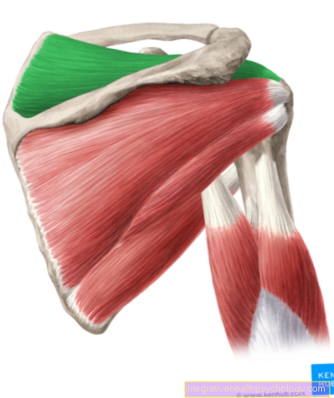
Shoulder and knee in an open MRI
Besides the Soft tissue representation and internal organs the open MRI is also used for diagnostic purposes Joint imaging. Especially that pool, the shoulder and the knee can be imaged in high resolution using the open MRI.
When the closed tube is used, it can often be observed that the illustrations show Creation of motion artifacts comes. The reason for this is mostly the fact that the position in which the shoulder to be examined or the knee to be imaged must be held during the examination is for the patient very uncomfortable is.
When taking pictures of the shoulder or knee, an open MRI has the advantage that the joint to be examined gently positioned can be. In this way, movement artifacts on the cross-sectional images of the shoulder and knee can be reduced.
A lower magnetic field strength is limited by the advantages that the image quality in an open system is usually worse than in a closed MRI.
Spine in an open MRI
A open MRI of the spine supplies overlay-free, high-resolution sectional images, the without radiation exposure can be made. In addition, the MRI scan of the Spine be performed in a comfortable position. Only in the MRI, regardless of whether it is a closed tube or an open MRI, can cartilage-, tape-, and Spinal cord structures the spine can be imaged in sufficiently high quality and resolution.
When examining the spine in an open MRI, the patient is pushed into the device either on their stomach or on their back. The examination of the spine in an open MRI usually takes between 15 and 30 minutes.
Among the most common Suspected diagnoses, which make it necessary to produce MRI cross-sectional images of the spine, include:
- disc prolapse
- Malformations of the spine
- Fractures (breaks) of the spine
- Inflammation
- Tumors
- Vortex sliding
Contrast media
By the gift of one Contrast media while performing an open MRI, a artificial density difference can be generated between different structures. A contrast agent is always necessary when very similar body tissues, for example Muscles and Blood vessels, should be separated from each other.
Even with an open MRI two types of contrast media a distinction can be made: iodine- and Contrast media containing barium sulfate. A contrast agent for performing an open MRI examination, which is a Iodine content contains, serves primarily to do so Kidneys, Vessels and internal organs to contrast sharper.
A contrast agent with Barium sulfate-proportion of however, is particularly suitable for sharper representation of the Gastrointestinal tract.
Read more on this topic at: MRI with contrast agent
Further topics
The following articles on the topic Imaging and MRI you might also be interested in:
- article MRI
- MRI examination
- You are here: Open MRI
- MRI thoracic spine
- MRI lumbar spine
- MRI shoulder joint
- MRI elbow
- MRI hand
- MRI heart
- MRI hip
- MRI abdomen
- MRI pregnancy
- MRI costs
- MRI knee
- MRI claustrophobia
- roentgen
- Computed Tomography
- Ultrasonic
- MRT in
For more diagnostic procedures, see Diagnostics A-Z.