Ribs
Synonyms
Medical: Costa vertebralis, Costae vertebrales
English: costal, ribs
introduction
The ribs as a whole make up the rib cage (thorax). Two ribs each are connected by the spine and the sternum.
Most people have 12 pairs of ribs (the number of ribs can vary), all of which are connected to our thoracic spine and determine the shape of the rib cage.
The upper 10 ribs (true and false ribs, see below) are also articulated to the sternum, the lower two ribs (rudimentary ribs, see below) are free.

Anatomy of the ribs
These 12 ribs (costae) are divided into three groups:
- True ribs (1st - 7th rib = costae verae)
- False ribs (8th - 10th rib = Costae spuriae)
- Rudimentary ribs (11th and 12th rib = Costae fluctuantes)
All ribs consist of a bony and a cartilaginous part. The ribs arise from the spine and become cartilaginous towards the end of the sternum.
The first rib is short and wide, protecting the chest from above. It is largely covered by the collarbone (clavicle).
The 8th - 10th ribs are called false ribs, because they do not reach the breastbone (sternum) directly, but instead merge with the 7th rib in a cartilaginous manner. The fused ribs towards the sternum are also known as the costal arch.
The 11th and 12th ribs are only stubby and do not end at the costal arch (rudimentary ribs).
In approx. 0.5% of all people, the 5th, 6th and 7th cervical vertebrae are mostly insignificant and have a so-called cervical rib, which is often only discovered by chance. In rare cases, a lumbar rib can also be formed on the first lumbar vertebral body, but often only as a stubby extension of the transverse process of the vertebral body.
Read more about this: Costal arch anatomy and diseases
Figure ribs

I-XII ribs 1-12 -
Costa I-XII
(I-VII) True ribs -
Costae verae
(VIII-X) false ribs -
Costae spuriae
(XI-XII) rudimentary ribs -
Cost. fluctuantes
- Rib head - Caput costae
- Rib neck - Collun costae
- Rib humps -
Tuberculum costae - Rib body - Corpus costae
- Rib head joint -
Articulatio capitis costae - Sternum - sternum
- Vertebral bodies -
Corpus vertebrae - Costal cartilage -
Cartilago costalis - Rib-sternum joint -
(Sternocostal joint) - Rib-vertebral body joint -
(= Point 5.)
You can find an overview of all Dr-Gumpert images at: medical illustrations
Which rib joints are there?
So that the ribs can move with the breathing movements, there are rib joints on the sternum and on the spine:
- Rib-vertebral joints
- Rib-sternum joints
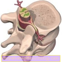
With the vertebral bodies of the spinal column, the bony end of the rib forms the costal vertebral joints (Costovertebral Joints), so-called ball joints. The round head of the rib lies in a depression in the vertebral body. The joints between the sternum and rib are called the rib-sternum joints (Sternocostal joints) designated. The first pair of ribs on the sternum handle (manubrium sterni) and the 2nd-7th. Rib on the body of the sternum.
The space between two ribs is called the intercostal space. The intercostal muscles are located here. In addition, nerves and vessels run along the inner underside of the ribs.

Figure chest (thorax)
- Collarbone (clavicle)
- Breastbone (sternum)
- Ribs (costae)
What is the costal cartilage?
The costal cartilage is physiological and a part of our rib cage. Anatomically, the costal cartilage connects our bony rib bodies (corpus costae) with the breastbone (sternum). As a result, the costal cartilage is located on the front side of the sternum. We have a total of twelve pairs of ribs. The first seven pairs of ribs are connected directly to the sternum via the costal cartilage. For this reason they are also known as "true ribs" (Costae verae).
The next three pairs of ribs (8th to 10th pair of ribs) are not individually connected to the breastbone like the “true ribs”, but rather they are cartilaginously attached to the respectively higher pair of ribs. This explains the term "false ribs" (Costae spuriae).
The last two pairs of ribs have no connection to the sternum, so that they are also known as rudimentary ribs (Costae fluctuantes). The costal cartilage is of particular importance for the elasticity of the chest, which is very important for breathing. Due to the cartilaginous connection between the ribs and the breastbone, the chest area can expand during inhalation, but it can also become smaller again during exhalation.
You can read detailed information on this topic here:
- Costal cartilage
- Which diseases of the costal cartilage are there?
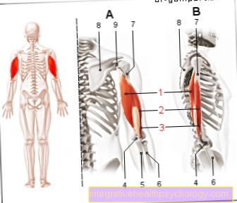
What are the rib muscles?
There are several rib muscles that play an important role, especially in breathing.
The intercostal muscles represent a large muscle group, which is made up of several muscles. On the one hand there are the outer, inner and innermost intercostal muscles (Musculi intercostales externi, interni and intimi), which stretch like a herringbone between the individual ribs. Their task is to widen the chest during inhalation (inspiration) and to reduce it during exhalation (expiration). On the other hand, the subcostal muscles (Musculi subcostales) lie under the ribs. These also belong to the intercostal muscles and are split off from the internal intercostal muscles. As a result, they also lower the ribs, helping with exhalation. Another muscle that belongs to the group of intercostal muscles is the transversus thoracis muscle. This tightens the cartilaginous costal arch and increases the resistance during inhalation.
There are also auxiliary breathing muscles that support the intercostal muscles when necessary. This includes a number of muscles, such as the pectoralis major and minor muscles, the serratus anterior and posterior superior muscles. Overall, these support inhalation. The different abdominal muscles are increasingly used when exhaling.
You might also be interested in: Torn muscle fiber between the ribs
Function of the ribs
The ribs determine the shape of the rib cage (thorax) and protect the lungs and heart.
In addition, the ribs are particularly important in our breathing, as they raise and lower the chest. This takes place in interaction with the two rib joints (rib sternum joints and costal vertebral joints) via various muscle groups that can be summarized as intercostal muscles. These are stretched between the ribs. The breathing movement between maximum inhalation and exhalation is usually more than 2 cm.
Cervical rib
A cervical rib is an accessory rib that attaches to a cervical vertebra.
Actually, humans have 12 ribs on both sides, which only arise from the thoracic vertebrae. If a cervical rib forms, it usually has no disease value. Most of the cervical ribs arise from the seventh cervical vertebra and are only weakly developed. They are mostly blunt and only cartilaginous or bound to the sternum with connective tissue. The incidence of cervical vertebrae is around one percent.
Neck ribs can also cause symptoms, depending on their size. In the vicinity of the seventh cervical vertebra, nerves and vessels run through the so-called scalenus gap. If the cervical rib becomes so large that it exerts pressure on this scalene gap, cervical rib syndrome can occur: Pressure on the nerves can lead to numbness or pain in the arm. If the artery is partially or completely squeezed off by the cervical rib, this can lead to reduced blood flow to the arm or to subclavian steal syndrome.
The therapy of a neck rib consists in the surgical removal.
This clinical picture can also be caused by a rib in the neck: Thoracic Outlet Syndrome
Which ribs can be removed for cosmetic reasons?
For cosmetic reasons it is possible to have some ribs removed. The rudimentary pairs of ribs (11th and 12th pair of ribs) are surgically removed. These have no connection to the breastbone (sternum) and are not involved in the structure of the costal arch. The aim of the patient is to have a wasp waist that should be as small as possible (up to 47cm). In Germany, this operation is not officially carried out because it represents a great risk and life with a wasp waist is dangerous.
On the one hand, the procedure is risky in itself, as the diaphragm and the lungs, for example, can be injured during the operation. On the other hand, living with fewer than 12 pairs of ribs is dangerous and has some disadvantages. The organs such as the spleen and liver lack important protection. This means that they are unprotected and, for example, are injured faster and worse in a fall. In addition, it is difficult to exercise with the ribs absent, as the upper body lacks overall stability.
You can find an overview of cosmetic surgery here.
Diseases of the ribs
Rib pain
Pain in the costal arch is a non-specific symptom and can have different causes. First of all, the patient should characterize the pain more precisely. Is it, for example, a pressure pain or a stabbing pain stimulus? Does the pain get worse when you exercise or when you breathe in? Based on this information, the attending physician can better assess the symptoms and the possible cause.
A bruised rib, for example, is a possible cause. This is indicated by a previous fall and bruises in this area. Then there is usually not only pain in the ribs, but also tension and bruises (Hematomas) in the area of the ribs. Especially with older patients or with particularly bad falls, it should be remembered that the ribs can also break. In this case, see a doctor as soon as possible, as the ribs are located above the heart and lungs and should therefore not be injured under any circumstances!
Inflammation of the pleurisy (pleurisy) can also cause pain in the costal arch. This is an inflammation of the pleura, which can be bacterial or viral. A characteristic of this disease is the increase in pain during inhalation.
In addition, it should always be considered whether the pain in the costal arch cannot also have organic causes. For example, the liver, gall bladder and also the stomach are possible. These organs are located in close proximity to the costal arch, so that the symptoms can be felt up to the costal arch.
Furthermore, muscular problems, such as muscle strains or torn muscles, can lead to pain. If there is tension or muscle soreness in the area of these ribs (for example intercostal muscles or serratus muscles), the patient may have the subjective feeling that the pain is coming from the ribs themselves.
If the pain comes from the muscles around the ribs, it is sufficient to wait two days, as the pain will then go away on its own.
Read more on the topic:
- Broken ribs or bruised ribs
- Broken rib pain
Projected pain on the ribs
However, there is also the possibility that the pain in the ribs is projected pain from the individual organs. Pain in the left costal arch can be associated with discomfort in the stomach, which is just below the lower left costal arch. Since the spleen is also located in the area of the lower left ribs, spleen complaints can also lead to pain in the area of the left ribs, although this is rare.
However, it is important to precisely characterize the pain. If the pain occurs suddenly and violently and if there is pain and tingling in the left arm in addition to the pain in the (left) ribs, a heart attack should always be considered! The emergency doctor must be notified immediately.
If pain in the right ribs is a problem with the liver or gallbladder. In this case, the pain in the ribs is accompanied by nausea or a bloated stomach.
If rib pain occurs when you cough, pleurisy (pleurisy) may be the cause. The duration of pleurisy can vary greatly. This can particularly affect patients with an existing lung disease.
Other rare causes of pain in the ribs include:
- Intercostal neuralgia
- Tietze Syndrome
- ankylosing spondylitis
Intercostal neuralgia
Intercostal neuralgia, also known as intercostal neuralgia, is razor-sharp pain, especially between the ribs, which is often caused by pinching the "rib nerves" in the thoracic spine.
Tietze syndrome is mainly characterized by pain in the area between the ribs and the sternum. This leads to inflammation of the cartilaginous joints that connect the ribs to the sternum (sternocostal joints).
Bechterew's disease (ankylosing spondylitis) is a rheumatic disease that is accompanied by inflammation, ossification and thus pain in the spine and ribs.
Read more on the topic:
- Rib pain
- Rib Block - How Can It Be Solved?
Broken rib
Ribs (Costae) form the outer shape of the thorax and are usually easily palpable and visible externally. Because of this unprotected position directly under the skin (or fat tissue), broken ribs (rib fractures) are not uncommon.
If the ribs are broken, this can usually be seen from the outside. Ribs can be broken, especially when falling or beating heavily in the chest area. Before the rib breaks, the ribs usually jump out of the joints that connect the ribs and breastbone (sternum) with one another (Articulationes sternocostales). Depending on the injury, not only one rib is broken, but several ribs at the same time.
In addition, a rib can break simply, i.e. only once, or it can be broken several times. In this case, one speaks of a row of ribs fracture. In general, strong external force is required to break a rib. If a rib fracture occurs spontaneously, one should immediately think of bone diseases such as osteoporosis. The proof of a broken rib can usually already be provided on the basis of the inspection, during which the doctor only looks at the patient and can thus already see the protruding rib. In addition, the doctor can feel the rib (palpation) to localize the break point more precisely. If the findings are unclear, an X-ray can also be taken.
It is always important to think about the surrounding structures when you have a broken rib. A broken rib itself is usually not a problem. However, it is fatal if, for example in a traffic accident, the broken rib damages the pleural leaves (pleural leaves) and air enters the pleural space. This phenomenon is known as pneumothorax and is associated with a collapse of the affected lung. This causes chest pain and shortness of breath. Damage to the spleen or the pericardium is also feared with a broken rib.
Usually, however, a broken rib is unproblematic and the broken rib does not have to be plastered, nor does a plate or screw have to serve as a stabilizer. Rather, conservative treatment with a three-week rest period is sufficient.
Read more on this topic at:
- Broken rib
- Chest x-ray (chest x-ray)
Bruised ribs
In the event of external traumatic influences (for example, a strong blow to the chest or a fall), the superficial position of the ribs can easily lead to rib bruises (Rib contusion) come.
It is important that a bruise does not exceed the elasticity of the rib bone. As soon as the elasticity is exceeded, a rib fracture occurs instead of a rib bruise (fracture). Since blood vessels and the rib nerves (Intercostal nerves) run, in addition to bruising the ribs, the small vessels can also burst. This then leads to an outflow of blood, which can be seen on the surface as reddening.
In addition, there may be a sensory disorder on the skin in the area of the bruised ribs (Loss of sensitivity). These sensory disturbances come about because superficial nerves have been injured or damaged and can no longer adequately convey their information to the brain via touches on the skin. Like the bruised ribs and the hematoma, this sensory disorder is uncomfortable but disappears again.
However, the consequences of a bruised rib can also be felt in the form of coughing, shortness of breath (dyspnoea) or severe pain in the area of the ribs or in the entire area of the upper abdomen. Especially if there is more pressure in the area of the bruise (for example the seat belt in a car), this can lead to increased pain.
If the pain becomes too severe, the patient can take pain medication. This is especially important if the patient has the feeling that his breathing has changed due to the pain or that he is adopting relieving postures that in the long run put a strain on the back. However, it is also important to always have a rib fracture clarified by a doctor. Older patients in particular easily mistake a broken rib for a bruised rib.
Further measures to alleviate the pain of a rib fracture are, on the one hand, adequate posture, and on the other hand, the patient can use cool compresses. These have the positive side effect that they cause the blood vessels to contract (Vasoconstriction). This reduces the leakage of blood and prevents too large a hematoma in the area of the rib fracture. Of course, this effect only occurs if the cooling compresses are placed on the corresponding area immediately after the rib bruise. If the ribs are severely bruised, an additional x-ray is recommended.
If the doctor is unsure whether soft tissue has been injured, he can also request an ultrasound.
Read more on this topic at:
- Bruised ribs
- Chest x-ray (chest x-ray)
What is pleurisy?
Inflammation of the pleura, also called pleurisy, is inflammation of the pleura (pleura). The pleura covers both the lungs and the chest cavity from the inside. This creates the so-called pleural space, which contains around 5 ml of pleural fluid. This ensures that breathing can take place with as little friction as possible.
Pleurisy is an inflammatory reaction that can have various causes. On the one hand, viruses, bacteria or fungi can cause pleurisy. These pathogens are usually absorbed via the airways and reach the pleura via the lungs. On the other hand, pleurisy is a concomitant disease (comorbidity) in various clinical pictures. Particularly noteworthy here are pneumonia, pulmonary embolism or pancreatitis (inflammation of the pancreas).
Breath-dependent pain in the rib area is characteristic of pleurisy. In addition, one can differentiate between dry and wet pleurisy. With dry pleurisy, only the pleura is inflamed, so that the patient feels very severe pain when inhaling. In contrast, the patient with wet pleurisy also has breathing problems. These are caused by the fact that more pleural fluid is produced, which accumulates in the pleural space. This compresses the lungs and makes it more difficult for the patient to breathe.
More information can be found here:
- pleurisy
- Duration of pleurisy
What is a rib bruise?
A rib bruise or bruise can be the result of trauma, such as a collision or a traffic accident. The soft tissue is crushed by a strong impact or a violent fall. The soft tissue includes, for example, the muscles that surround the ribs as well as the lungs themselves. The squeezing injures small blood vessels, causing small bleeding into the tissue.
Similar to a bruise (hematoma), this injury is harmless, although it can be very painful. A bruised rib is often expressed as a bluish discoloration in the area of pain. The attending physician can rule out a broken rib with an X-ray. The bruised ribs usually heal on their own after a few days. The patient should take a few weeks of physical rest and refrain from particularly physical sports such as football or martial arts for a few weeks. If the ribs are particularly badly bruised, supportive physiotherapy can be carried out.
Inflammation of the rib joint
The ribs (costae) form the outer shape of the thorax and are important bones that support breathing. There is no inflammation of the ribs themselves. However, the joint that connects the ribs to the breastbone (sternum) can become inflamed. This is the so-called Tietze syndrome, in which the costal cartilage that attaches to the sternum is inflamed. There is severe pain in the chest area.
The causes of Tietze syndrome and the associated inflammation of the ribs are not yet known. However, since only the first 7 ribs are "real" ribs (costae verae) and are connected to the sternum by cartilage, the inflammation only occurs in the first 7 rib joints (sternocostal joints). Most often, however, inflammations occur in the 2nd to 5th grade. Rib in front.
In addition to the inflammation of the costal cartilage, pain in the area of the ribs can also occur after an inflammation caused by herpes zoster (shingles). The reason for this is a nerve irritation (neuralgia) in the area of the ribs, which is caused by the inflammation. In this case, however, the ribs are not directly affected by the inflammation, rather it is an irritation of the rib nerves (= intercostal nerves, i.e. intercostal neuralgia) from the inflammation with the virus.
Read more on this topic at: Tietze Syndrome
Adjust ribs
The ribs, together with the spine and sternum, form the bony security for our upper body (thorax) and envelop the heart, both lungs, spleen and kidneys. Athletes in particular often have the problem that they “dislocate” their ribs if they move incorrectly. These are shifts in the area of the costal vertebral joints, which can then lead to severe pain and possibly even shortness of breath.
It is now important not to try to adjust the ribs yourself. On the one hand one can only worsen the situation if one is ignorant, on the other hand a dislocated rib is already painful enough. Adjusting the rib by yourself without specialist knowledge is therefore not desirable. Osteopaths, physiotherapists or, in some mild cases, the family doctor are recommended. In order to counteract a renewed dislocation of the ribs, one should also pay more attention to sporting activity (especially back muscle training). Massages, however, are not beneficial. Since athletes in particular often suffer from displacements in the area of the costal vertebral joints, they can have their osteopath or physiotherapist show them how they can straighten their ribs themselves.
One exercise that must be discussed beforehand with the respective physiotherapist is that the patient lies down on the non-painful side, tucks a towel under the lying side at the level of the painful rib and slowly breathes deeper and deeper against the pain. During this exercise, the ribs should be mobilized more. However, if the pain worsens, stop doing the exercise and see a physiotherapist again so that he can professionally adjust the rib.
You might also be interested in: physical therapy