Femoral neck
introduction
The thigh bone (also: femur) is the longest bone in humans and creates a connection between the pelvis and the lower leg bone. It is connected to the other bones through the hip or knee joint. At the end of the hip, the thigh bone has a spherical shape, which is why it is called the femoral head (also: caput femoris). The end that points towards the knee has more of a fork-shaped structure and ends in two cylindrical structures, the articular knot (also: epicondylus medialis and epicondylus lateralis)

The thigh bone consists of two parts of different lengths, which is why one can also speak of an L-shape. The shorter piece points towards the hip joint and the longer towards the knee. The connection between the short and the longer part is called the femoral neck (also: collum femoris). Due to the effects of physical force, this area is particularly at risk for broken bones.
Also read more on the topic: Femoral neck fracture
It is important to mention the two rolling hills (also: trochanter major and trochanter minor), which are located directly at the transition from the femoral neck to the long part of the thigh bone (also: corpus femoris). These bumpy structures form the attachment point for many muscles in this area. The same function also has a rough, linear elevation (also: Linea aspera) on the back of the long part of the thigh bone.
Functionally, the thigh bone has to bear the weight of the human body on the one hand, whereby it has to withstand both compressive and tensile stresses. On the other hand, it must allow a certain mobility in relation to the joint surfaces of the adjacent bones in order to enable the lower extremity to perform more functions. All in all, a versatile task.
Waistline of the femoral neck
"Waist" means the slimming of a structure at a certain point. In connection with the femoral neck, this means the anatomically conditioned thinning of the femoral neck compared to the rest of the thigh bone or the long portion of the thigh bone. The waist at this point allows a greater range of motion for the thigh, but above all better flexion and rotation in the hip joint. If, on the other hand, the sidecut is not sufficiently pronounced, for example due to a congenital form disorder at this point, this can lead to the clinical picture of CAM impingement. These patients then mainly have problems with flexion in the hip joint.
Also read more on the topic: Hip impingement
Femoral neck angle
The angle between the longitudinal axis of the femoral neck (also: collum femoris) and the longitudinal axis of the long part of the femur (also: diaphysis) is called the femoral neck angle. Alternatively, the term des CCD angle (Center-Collum-Diaphyseal angle) is used. This should ideally in healthy adults 126° be. If this is the case, one speaks of a physiological situation (also: Coxa norma), in which the bone-specific structures for pressure and tensile loads (also: pressure and tensile trabecula) are precisely balanced.
However, there are also deviations in the CCD angle, each of which can have different consequences for bone structure and possibly even for joint function.
Is the angle for example too small (for example smaller than 120 °) results in a increased tensile load due to increased bending stress. This leads to increased formation of draw trabeculae and is called Coxa vara designated.
Is the angle on the other hand too large (for example greater than 130 °) is the Increased compressive stress and it comes to compensatory increased formation of pressure trabeculae. If this is the case, one speaks of one Coxa valga.
Thigh neck pain
If the thigh bone is misaligned, the body automatically attempts to compensate for this. As a result, the person affected may not feel any discomfort in the acute situation, but this will lead to signs of wear and tear (for example Hip osteoarthritis) and improper stress with pain symptoms. It is therefore important to diagnose misalignments at an early stage and, if necessary, to have them corrected in order to prevent chronic consequences and further pain as far as possible.
Also read more on the topic: Hip pain
Femoral neck diseases
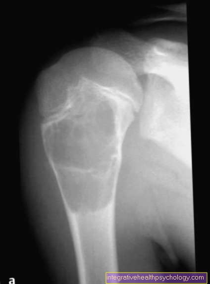
Femoral neck fracture
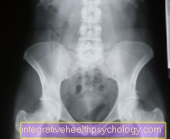
A femoral neck fracture is a fracture of the bone between the shaft and the head of the femur. Because the bone is angled at this point, it is easier to break if you fall.
Older people over the age of 60, and especially older women, have an increased risk of breaking the femur as a result of a fall. The bone loses its stability with increasing age, since the bone is broken down and consequently a comparatively light fall can lead to a break.
In younger people, femoral neck fractures usually occur as a result of high-speed trauma, for example when riding a motorcycle or as a result of traffic accidents. Different break locations are distinguished depending on the force applied. Those affected usually report severe pain when moving the leg. Often the corresponding leg is turned outwards and slightly shortened when lying down.
Depending on the type and extent of the fracture, treatment is either surgical or immobilization for some time and subsequent gradual movement is recommended.
Read more detailed information on the subject of femoral neck fracture here.
CAM impingement
An impingement is generally a bottleneck between two structures. Translated it means "collision". CAM impingement involves a narrow point between the head of the femur and the acetabulum. The bottleneck is caused by an insufficiently developed waist at the transition from the femoral head to the femoral neck or by a bony attachment.
The fact that the bone does not lose its circumference at the transition to the femoral neck means that it repeatedly hits the socket when the hip moves. The cartilage can be injured and torn or inflamed by this constant irritation, especially during exercise.
CAM impingement manifests itself as severe pain in the groin when standing, walking or sitting for long periods of time. Usually there is restricted movement in the hip joint.
CAM impingement mostly affects young men from the age of 20. If such complaints are noticed, a doctor should be consulted. If left untreated, osteoarthritis (joint wear) of the hip joint occurs in most cases. Surgical or conservative therapy is available as treatment options, which should be weighed up by an appropriate specialist depending on the severity of the impingement.


.jpg)