Spondylolisthesis
Synonyms
Spondylolisthesis, vertebral sliding, vertebral body sliding, sliding vertebrae, degenerative spondylolistesis, degenerative spondylolistesis, congenital spondylolistesis, congenital spondylolistesis, back pain
Definition of spondylolisthesis
A Spondylolisthesis / Spondylolisthesis denotes a Vertebral body sliding. Almost always it is Lumbar spine affected. There are innate and acquired forms of Spondylolisthesis known.

Of the common causes of spondylolisthesis, a child / adolescent form can be attributed to wear-related (degenerative) distinguish adult form. In the child / adolescent form, there is an interruption of the vertebral arch (Spondylolysis) to instability of the vertebral bodies among one another. The lowest part of the vertebral body (segment) of the lumbar spine, lumbar vertebrae 5 to sacral body 1 (short: L5 / S1). Here the vertebral arch of L5 is diseased and can be accessed via the Sacrum (Sacrum) slide forward towards the abdomen (isthmic form of spondylolisthesis).
The adult form of spondylolisthesis is part of the degenerative spinal diseases / back problems. The vertebral body section lumbar vertebrae L4 to lumbar vertebrae L5 is particularly affected. There is no lysis zone (interruption of the vertebral arches). The cause of the vertebral slipping (spondylolisthesis) is a degenerative instability due to a decrease in height of the Intervertebral disc between L4 and L5 as well as general loosening of the stabilizing segment structures (ligaments, muscles, etc.).
Appointment with a back specialist?

I would be happy to advise you!
Who am I?
My name is I am a specialist in orthopedics and the founder of .
Various television programs and print media report regularly about my work. On HR television you can see me every 6 weeks live on "Hallo Hessen".
But now enough is indicated ;-)
The spine is difficult to treat. On the one hand it is exposed to high mechanical loads, on the other hand it has great mobility.
The treatment of the spine (e.g. herniated disc, facet syndrome, foramen stenosis, etc.) therefore requires a lot of experience.
I focus on a wide variety of diseases of the spine.
The aim of any treatment is treatment without surgery.
Which therapy achieves the best results in the long term can only be determined after looking at all of the information (Examination, X-ray, ultrasound, MRI, etc.) be assessed.
You can find me in:
- - your orthopedic surgeon
14
Directly to the online appointment arrangement
Unfortunately, it is currently only possible to make an appointment with private health insurers. I hope for your understanding!
Further information about myself can be found at
Spondylolisthesis vera
Spondylosthesis vera or 'real spondylolisthesis‘Refers to the sliding or sliding of a vertebra forwards, probably due to a congenital disorder. With this clinical picture there is a faulty formation of the vertebral arches (Spondylolysis) during development in the womb. The exact causes of this malformation (Dysplasia) are still unknown. Theories look for the reason in the evolution of the upright gait when humans rose from the quadruped position on their two legs.
Thus the spondylolisthesis vera clearly shows vertebral displacements without gaps in the Vertebral arch delimit. These include degenerative spondylolisthesis (Pseudospondylolisthesis), Retrolisthesis (Backward vortex sliding), rotating sliding and vortex sliding as a result of osteoporosis, tumor, Inflammation, rupture, etc.
At the beginning of spondylolisthesis vera, the defect is in the vertebral arch. Most of the time this is not made entirely of bone, but rather by a connecting piece made of softer material, such as cartilage, interrupted. It is not uncommon for the faulty piece to be extended. Due to the increasing stress on the back, the soft cartilage parts dissolve over time. The interruption of the arch is usually found in the area between the upper and lower articular process (Interarticular portion) as well as on both sides. So the stability in the spine is no longer guaranteed. As a result, the corresponding vertebral body slides forward (ventrally = ventral), including the part of the Spine.
In many cases, the spondylolisthesis vera runs smoothly. Typically it happens by chance in young adulthood as part of a X-ray examination discovered. It is relatively widespread among the population (2-4%)!
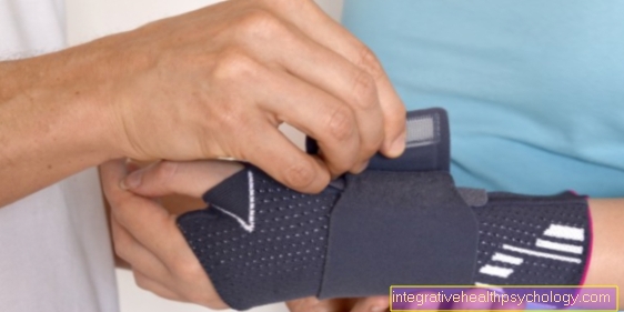
In children, however, there is a risk of further, massive slippage of the vertebrae as they grow, so that strong ones Back pain up to neurological deficits are possible. If painful spondylolisthesis vera is confirmed, symptomatic pain medication can be administered. In addition, the abdominal and back muscles should be through physiotherapy be strengthened. A support bandage or a corset can also help.
However, it should be emphasized that spondylolisthesis is often free of symptoms! So as long as there is no pain, treatment is not necessary.
Symptoms of spondylolisthesis
The symptoms that can be caused by spondylolysis / spondylolisthesis are diverse, not very characteristic and depend on the severity of the disease.
Most patients with juvenile spondylolysis / spondylolisthesis have no symptoms. The diagnosis is often an incidental radiographic finding. Degenerative spondylolistheses can also be silent, but are more likely to cause problems due to the general signs of wear and tear on the spine.
Symptoms of spondylolysis / spondylolisthesis may include:
- Back pain (lumbago) at rest, when moving, when exercising
- Pain in the back radiating to the legs (lumbar sciatica), either according to the area of spread (dermatome) of a nerve root or unspecific (in the case of additional degenerative diseases or advanced vertebral sliding).
- Sensory disturbances (hypoesthesia), paresthesia of the legs (with advanced vertebral sliding).
- Leg weakness
- Restricted movement of the lumbar spine
- Hip lumbar stiffness
- Muscle tension
- Bladder / rectal disorders (with advanced vertebral sliding with spinal cord narrowing).
Neurological deficits (paralysis, sensory disturbances, bladder - rectal disorders) are reserved for very advanced cases of spondylolisthesis and are rather the exception because the neural structures (spinal cord, nerve roots) adhere to the changed spatial conditions, with the usually slow process of vertebral sliding up to one can adjust to a certain extent.
Spondylolisthesis can also trigger a so-called cauda equina syndrome, which should be recognized urgently and, if left untreated, can lead to permanent paraplegia. We therefore recommend our website for further information: Cauda equina syndrome - do I have paraplegia?
frequency
The rate of spondylolysis in adults is 6%, in children up to 6 years of age 4,4%.
In about 80% of the spondylolysis there is a disease of the 5th lumbar vertebra. The 5th lumbar vertebral body slips forward. In about 20% there is only a unilateral spondylolysis defect in the vertebral arch.
Boys are 2-3 times more likely to have spondylolysis. However, severe vertebral sliding occurs 4 times more often in girls.
In the majority of cases, vertebral slippage occurs as a result of spondylolysis between the ages of 12-17. Year of life. After growth is complete, the spondylolistesis is no longer expected to progress.
Degenerative spondylodesis affect the L4 / 5 segment in approx. 80%, L5 / S1 in approx. 15% and rarely the upper and middle lumbar spine segments.
10% of women over 60 years have degenerative spondylolisthesis. Vertebral slippage over 30% of the vertebral body's longitudinal diameter is rare.
Classification according to Meyerding
Information on the Meyerding classification
The severity classification according to Meyerding, which is based on the Extent of the tilt angle of the two vertebrae justified to each other. For this you need a lateral X-ray image of the spine, which is part of the standard diagnosis of spondylolisthesis.
The classification according to Meyerding is different 4 degrees of severity the spondylolisthesis. There are sometimes different descriptions for assessing the graduation.
The lower vertebral body of the two vertebrae sliding together is divided into 4 parts. If the upper vertebra is shifted by less than ¼ in relation to the lower one, it is called a Meyerding grade I. If the sliding process has progressed further, namely by up to 50%, it is a Meyerding grade II. An offset of 50 up to 75% is a Meyerding grade III. An offset of over 75% defines a Meyerding grade IV. According to some authors, there is also a Meyerding grade V in some cases. This is what is known as spondyloptosis, in which the two vertebrae no longer have any contact with one another. In the strict sense of the word, grade V does not represent a spondylolisthesis.
Grade 1
A spondylolisthesis grade I according to Meyerding is the morphologically most easily pronounced form of vertebral sliding. The offset between the vertebrae is less than 25%, which means that the vertebrae are displaced from one another by less than ¼ of the width of the lower vertebral body. You can see this on the side of the X-ray image. The extent of the vertebral slipping can, but does not have to, correlate with the extent of the symptoms. 90% of the spondylolisthesis is asymptomatic anyway. In combination with other diseases such as spinal stenosis, however, it can cause symptoms. Therapy is not absolutely necessary. Physiotherapy and good strengthening of the back muscles can prevent further vertebral slipping.
Grade 2
A Meyerding grade II spondylolisthesis is characterized by an offset of the two vertebrae between 25 and 50% to each other.
The displacement of the sliding vertebrae is determined on the basis of a side view in the X-ray and says something about the extent of the vertebral sliding. The graduation does not capture the symptoms. In over 90% of the cases even show up no symptoms. Therefore, a grade 2 according to Meyerding is a conservative therapy in the form of physiotherapy and follow-up monitoring is sufficient. Trigger sports such as gymnastics or weight lifting should be avoided. The Follow-up is important insofar as it is more likely that young patients will have a progressive course, i.e. rapid progression of the vertebral sliding, than older patients. In the latter, spondylolisthesis usually occurs as part of degenerative changes in the spine.
Grade 3
If the vortex is offset 50 to 75% to each other one speaks of a spondylolisthesis grade III according to Meyerding. This is a severe vortex slidingwhich can lead to spinal instability. A operative therapy can in this case considered to restore the stability of the spine. With a grade III according to Meyerding, symptoms are likely but not obligatory.
anatomy
The Lumbar spine (= LWS) turns off formed by the five lumbar vertebrae of the spine. Since they are located in the lower part of the spine, they have to carry the highest percentage of weight. For this reason they are also much thicker than the other vertebrae. However, this does not avoid the signs of wear and tear that occur very frequently, particularly in this area. For example, joint wear and tear are Herniated discs in the lumbar spine most frequently.
The lumbar spine also differs in structure from the other areas of the spine. For example, from the second lumbar vertebra onwards there is none Spinal cord more in front, but only individual nerve roots that pull further down and emerge from the nerve root holes intended for them. This area, in which the spinal cord ends and the spinal canal is filled with nerves, is known as the "horse's tail", or medically as Cauda equina.

X-ray image of the lumbar spine (lumbar spine)
- Intervertebral disc (blue)
- Vertebral bodies
- Sacrum (red)
The is stabilized Lumbar spine (LWS) through the intervertebral discs between the vertebral bodies and a complex system of ligaments and muscles.
Of particular importance for the clinical picture of children / adolescents Spinal fusion is the vertebral arch area between the upper and lower vertebral arch joint (Interarticular portion). In this area there is a zone of osseous loosening (lysis zone) vortex sliding can occur.
root cause
Spondylolisthesis can have various causes. A distinction is made between the following causes:
- Congenital, dysplastic spondylolisthesis (rare)
- Child / adolescent (isthmic) spondylolisthesis (common)
- Degenerative, adult spondylolisthesis (common)
- Post-traumatic spondylolisthesis (rare)
- Pathological spondylolisthesis (rare)
- Postoperative spondylolisthesis (rare)
Spondylolisthesis can be a cause of chronic both in childhood and adolescence as well as in adulthood Back pain be.
- more on the subject Spondylolisthesis emergence
diagnosis
Spondylolisthesis are mostly called Incidental finding in the x-ray discovered as they are asymptomatic in most cases. Likewise, they can also be made by the standard preparation of an X-ray image based on Back pain, possibly with Radiance in the legs or in combination with Sensory disturbances in the lower extremity, be diagnosed. If the spondylolisthesis is severe, a Trunk displacement, also as Ski jump phenomenon during the physical examination by the doctor. In any case, imaging examinations are, above all, that X-ray in two planes (from the side and from the rear), necessary to assess the extent of the disease. Since slipping of the vertebrae often only occurs with a certain posture or position of the person concerned, it is with Suspected spondylolisthesis useful, in addition Function recordings close. These are in this case X-rays while standing when leaning forward or leaning back.
There is then typically one on the X-ray Interruption of the bony vertebral arch (Spondylolysis) and a Sliding of the vertebral body to recognize backwards or forwards. Depending on the Spinal canal expanded or narrowed, which explains parts of the symptoms. Usually goes with the Displacement of the vertebral body also a degenerate, i.e. worn, Intervertebral disc hand in hand.
To make a more accurate assessment of the Spinal anatomy to enable an additional MRI (Magnetic resonance imaging) or CT (Computed tomography).
A CT is ideally suited to the Condition of the bony structures to be able to assess, whereas by means of a MRI the Discs and nerves considered particularly well can be.
therapy

For the Therapy of spondylolisthesis currently exists no valid guideline, which is why it must be treated according to the current study situation and / or personal experience of the doctor. In any case, this is important when deciding for or against certain therapy options Degree of vertebral sliding and whether it is a real spondylolisthesis (Slipping of a vertebra due to the formation of a gap in the vertebral arch) or a Pseudospondylolisthesis (the gliding of a vertebra despite intact vertebral arches).
Usually (also because of the unclear study situation) a physiotherapy treatment to Strengthening the back muscles and Reduction of the hollow back (Lordosis) aimed at. In addition, the Administration of pain medication. A Pain therapy by injecting Local anesthetics in the skin (Infiltration therapy) or directly to the Nerve root (Perdiradicular therapy) is also often used. Likewise can medical massages cause a reduction in pain. The administration of Muscle relaxants However, (drugs to relax the muscles) turned out to be not as effective. A regular one process control is necessary in any case.
Especially with children may practice certain sports such as gymnastics, javelin throwing or swimming (especially the technique of dolphin swimming) Trigger of spondylolisthesis be. If the disease has already occurred, that is too Avoid these high-risk sports Part of the therapy. Follow-up monitoring in children is also even more important than in adults, as they are at higher risk of severe disease. A special feature of children is that the spondylolisthesis heals in some cases through the Application of a plaster cast or a corset can be achieved.
The as the last option to consider is after all that operative treatment. This is mainly used when the pain cannot be managed conservatively (using the methods mentioned above) or more serious complications, such as Nerve damage occur. An attempt is made to use the Vertebral bodies returned to its physiological position and stiffened (Spinal fusion). This process ultimately leads to one irreversible loss of movement in the treated spinal column.
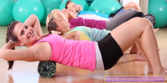
Training exercises
Usually spondylolisthesis does not cause any symptoms. It is asymptomatic and so many of those affected do not even notice it. However, some people experience pain and other ailments as part of their spondylolisthesis.
At a light spondylolisthesis it is recommended by targeted exercises to strengthen the back and abdominal muscles.
A couple of exercises should be explained here as examples:
1st exercise: Lie flat on your stomach. Tense your stomach and back. Now lift your upper body slightly. The view points downwards. Hold these positions with your arms extended forward for 10 to 15 seconds. If you want to intensify the exercise a bit, paddle your arms alternately. Repeat the exercise about 10 times.
2nd exercise: Lie with your back on the floor. Now embrace your knees with your arms and pull your chin towards your chest. Rock it back and forth and back and forth about 15 times. You can repeat these exercises 3 to 5 times.
3rd exercise: Lie on your back on the floor. Now put your legs up. Place your arms next to your body. Now lift your pelvis, shoulders and head stay on the floor. Your body (shoulder, pelvis and knees) forms a line. Hold this position for about 10 to 15 seconds. Then put the pelvis back down. Do 5 to 10 repetitions.
4th exercise: Lie on your side. Now support yourself on your right forearm. The legs are stretched out one above the other and the pelvis is raised. The legs and torso now form a line. Tense your stomach and back. Hold the position for 10 to 15 seconds. Then the side is changed. Do 10 to 15 repetitions per side.
Surgery (spinal fusion)
A surgery is always the last option to be considered if previous attempts at therapy have failed or were not even considered. It is particularly suitable if the pain cannot be managed conservatively, that is Vortex sliding increases very sharply in a short time or one Impairment of nerves occurs. This can be in addition to pain Urinary retention or one Fecal incontinence up to Muscle failure express. This tries to return the vertebral body to its physiological position and to stiffen what as Spinal fusion referred to as. In the best case scenario, this can result in a full load capacity of the spine are made possible.
To Start of the operation usually two Accesses to the spine created; a front (ventral) and a back (dorsal). Once the access points have been created, screws are inserted into the affected and adjacent vertebral bodies and with them Rods or metal plates connected together. Sometimes the space between the vertebrae is added Bone chips brought in. Special titanium cages can also be inserted into the intervertebral space, which support bone healing. After the operation, a Control X-ray prepared. Once all of the vertebrae have grown together, the spondylodesis material can be removed in another operation.
The Surgical technique of spinal fusion Depending on the length of the treated spinal column, leads to a complete, irreversible one Loss of mobility in this area, but the spine is fully resilient. Basically there is also the risk of Nerve damage and the Formation of scarswhich can sometimes cause very severe pain. These two complications are commonly called Failed back surgery syndrome designated.