forearm
definition
The forearm is the connection between the hand and the upper arm and is responsible for the transmission of force to the hand. It is the insertion of numerous muscles that move and control the hand itself.
In addition, it directs all vessels and nerves to the hand or to the body trunk and is also supplied by them itself. Just like the lower leg, the forearm is made up of two bones. These cooperate with each other and thus form two other important joints that allow the hand to rotate and are therefore essential for numerous movements.
Due to its superficial and often clearly visible veins, it is the preferred puncture point for blood withdrawals or intravenous injections.
Diseases: Pain in the forearm - what's behind it?
The forearm is formed by the ulna and radius, two almost parallel bones that are connected by a layer of connective tissue. There are many ligaments, tendons, and muscles in the forearm that can cause pain. Here is the Pain very variable, it can appear anywhere on the forearm, it can pulling, oppressive or throbbing be just under pressure occur or permanent to be available. A Broadcast into the hand or over the elbow to the upper arm are not uncommon.
As a rule, the pain results from excessive or improper strain on the forearm. This incorrect load can tense the muscles or cause sore muscles, the tendons can become irritated and inflamed.
Such forearm pains are widespread, so not a rarity. Typical diseases are tennis elbow and bursitis. With such an overstrain, it may be sufficient to take care of your arm and the to stop stressful activity.
However, it is also possible that the arm or ulna and / or spoke are broken after a fall. After a fall or accident that has resulted in an injury to the forearm, one usually suffers from acute pain. If so, a doctor should be consulted. However, it is just as important to see a doctor if you are suffering from chronic pain that does not get better even after taking care of your arm.
The forearm fracture
The forearm consists of two long bones, which can break when subjected to external force. Common causes are Falls on the arm or hand and injuries Accidents. The forearm fracture can get through Pain in the forearm, through a swelling and / or a Misalignment make noticeable of the arm. Often is a Move of the arm painful and can also possibly crunch. In the hospital, the fracture is usually diagnosed with two x-rays from different perspectives and an operation is decided based on how many parts of the bone are broken and whether a joint is involved.
With a forearm fracture, it is possible that there is an isolated fracture in which the ulna or radius is fractured (Broken) or that there is a combined fracture affecting both bones.
Often only the spoke breaks close to the hand (distal radius fracture), this fracture accounts for 25 percent of all bone fractures in adulthood. The cause of this fracture, which is almost always operated on, is a Fall on the hand with outstretched arm. The forearm fracture of both bones is less common and the isolated fracture of the ulna is extremely rare.
The fractures are usually operated on if the bone has shifted or the wrist is affected. If this is not the case, they grow back together well without outside help, so that no damage remains.
How do you wallpaper the forearm correctly?
Taping is still a very young form of therapy and is based on the experience of doctors, physiotherapists and athletes. For this reason, problems with the forearm should always be clarified with a doctor first and the taping should preferably be carried out with therapeutic guidance.
In sports, such as Climbing, tennis, volleyball, golf ect. the forearm is very stressed and strained, which is why taping is recommended. The tape relieves the ligaments and muscles and stabilizes the joint. This prevents overstressing of the forearm through prevention.
Before the so-called Kinesiology tape aIf the skin is glued on, it should be depilated and free of oils, fats and water. The tape is affixed about 30 minutes before exercise; It is important to pay attention to the correct tape size. It should be considered which sport is being performed in order to put the tape in the right place. The tape placements differ, whether the elbow or the wrist should be supported.
In addition, there are specific tape instructions for a golfer's arm and a tennis elbow.
- The golfer's arm shows pain on the inside of the elbow, but the tape can not only help with this, but also with problems of the forearm flexor muscles.
- With tennis elbow, on the other hand, the pain is localized on the outer side of the arm.
The tape helps not only tennis or badminton players with such pain, but also people in general who suffer from pain in the outer arm. When sticking the tape, it is important that the corners are rounded off with scissors and that the tape is pressed down well so that the hold is improved.
How can you train the forearm?
For most people, the strength that the muscles of the hand can exert is very weak, so that it is hardly possible to hold heavy weights. So if you want to lift heavier weights, do more pull-ups or prevent injuries to the forearm, hand strength and thus the forearm should also be trained.
There are isolated forearm exercises, but also exercises that train several parts of the body, including the forearm. Other exercises train grip strength - such as pull-ups or deadlifts.
- Forearm curls are a particularly good exercise for the forearm. This exercise trains the hand extensors, hand flexors, the long palm muscles and the deep finger flexors. Before starting the forearm curls, your arms should be warmed up to prevent injury. A dumbbell is required for the exercise. Sit on a bench or chair and place your hand with the dumbbell on your thigh so that your wrist and hand are above your knee in the air. The inside of the arm is at the top. The upper body is bent slightly forward and the back is straight. When inhaling, the hand is lowered and the dumbbell is brought down, while when exhaling, the wrist is bent and the dumbbell is brought up. This is an effective exercise for the forearm, but it should also be ensured that the uterine arm curls are reversed, i.e. with the top of the arm up. Thus, the wrist flexor inside and also the wrist extensor outside are trained evenly.
- A very simple but also effective exercise for the forearm is to squeeze an object, for example a kneading ball. When training, you should make sure that you often do short training units instead of several hours once a week.
What is a crutch?
A forearm crutch is also referred to as a crutch in everyday usage. If you suffer from an injury, such as a fracture of the lower extremity (leg or foot), the leg may no longer be able to carry your entire body weight without being damaged. A crutch is recommended as it shifts part of the body weight from the legs to the arms. This protects joints and bones.
This not only relieves the legs a little, but it is also possible that a safe gait is created if the person concerned previously suffers from an unstable gait pattern. The hand and forearm lie or are supported on a cuff. The cuff is available in a wide variety of designs so that the weight is evenly distributed and there are no pressure points on the hands. The contact surface is covered with plastic accordingly. The cuff ends in a support tube that leads to the floor and has a rubber knob there. The rubber prevents slipping on a slippery or wet floor.
As with the arm cuff, there are also different variations of the support tube, such as longer models for tall people. Different forearm crutches also differ in the weight they can carry. The average crutch is designed for approx. 100 kg, but there are also some for more body weight. As a rule, they are made of a light metal (aluminum) or made of plastic. In addition to the classic crutch, there are also special ones, such as the arthritis crutch.
If you are interested in the subject of "forearm crutches", read our next article at: Forearm crutch
The forearm bandage
An underarm bandage will also be used Forearm cuff or Sports bandage called. A bandage protects the forearm from overload and hypothermia during sports. However, it can not only be used as a preventive measure, but also if you have ever suffered from problems with your arm, such as a tennis elbow. It is also used for the conservative treatment of carpal tunnel syndrome.
In addition, the bandage protects against external influences in the Ball sports and can thus prevent injuries. A bandage is usually made of synthetic fibers that can be washed. The material is thin-walled and offers flexibility in the joint area. When exactly it makes sense to wear a bandage should be discussed with the doctor and depends very much on the patient's problem.
The forearm splint
A forearm splint is placed around the forearm and wrist so that the wrist is fixed and stabilized. It is recommended to wear a forearm splint if there is acute or chronic irritation to the wrist, if there is pain in the wrist, if you have carpal tunnel syndrome, or if you are immobilized after surgery or an accident.
The forearm splint immobilizes the wrist, finger mobility remains, so that the tweezer grip can also be performed with the thumb and forefinger. The splint ends in the lower third of the forearm. Depending on the indication, there are also splints that fix both the thumb and the rest of the fingers and thus limit their mobility. There are very different types of rails on the market, so that you can find a rail that is comfortable to wear. The splints are mostly worn at night to keep the wrist in the middle position (Zero position) keep calm, because the complaints are usually more frequent at night than during the day, as the hand bends while sleeping. All rails are available for both the left and the right hand. As a rule, they are made of allergy-tested material (plastic) and are easy to put on and take off using Velcro fasteners.
Swelling on the forearm
Swelling, also called edema, is a build-up of fluid in the tissue. The swelling can be easily seen from the outside, as the arm is thicker, so that otherwise matching pieces of jewelry or watches are very tight or even too small. If you press in the skin, dents appear. The skin in the area of the swelling is very tense, so that the wrist and / or the elbow joint can no longer be moved properly and a feeling of tension or even pain can occur in the swelling. These symptoms are a good way to diagnose edema.
The cause of the forearm swelling can be very different.
- It is possible that a harmless accumulation of water that goes away on its own triggered the swelling, for example during pregnancy or if you haven't moved your arm for a long time.
- Furthermore, some medications trigger edema as a side effect or the swellings can arise as a result of an allergic reaction.
- In addition, edema can occur as an accompanying symptom of a serious illness. An example of this is cardiac insufficiency (Heart failure) to call. The heart is unable to pump the blood through the body with enough force that it accumulates in the vessels in front of the heart and the veins are exposed to increased pressure. This causes fluid to move from the veins into the tissue, causing water to accumulate in the arm and leg.
- If the lymphatic system is damaged, edema can also occur in the forearms.
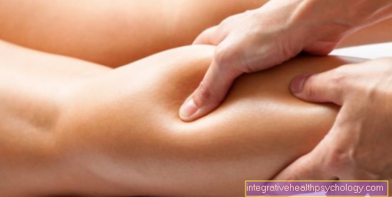
Muscle pain in the forearm
Muscle pain in the forearm can have a variety of causes, but most of the time it is due to overuse or improper use. A cramped arm posture, for example due to long computer work or the suddenly intensive sports program, are often responsible for sore muscles in the forearm and thus for pain there. If the pain occurs depending on such an activity, there is no need to worry, the arm should still be spared until improvement occurs.
Muscle pain is also a not uncommon symptom of the flu. If the muscle pain is chronic, a doctor should be consulted, as this could also be a muscle inflammation or soft tissue rheumatism.
Inflammation in the forearm
Inflammation in the forearm is very general; it can come in many different forms and have very different causes. It is possible that ligaments, eyes, bursae, or muscles are inflamed.
The typical triggers can be hypothermia of the arm, constant friction or pressure, an incorrect load and overload. A common misuse and overstrain arises from unfamiliar sport that is suddenly and intensively exercised or from prolonged computer work. In most cases, forearm inflammation is a reaction to a physical and / or mechanical stimulus.
If the muscles or tendons are inflamed, the following five symptoms occur, which describe a typical defense reaction of the body: The arm is reddened, swollen, sensitive to pain, warmed up and restricted in its function.
Bruise in the forearm
A bruise is an injury that can occur on any part of the body because it is caused by a blunt external force. Typical causes of a bruise on the forearm are a blow, impact, fall on the arm, or crushing of the arm. A bruise is a compression of the tissue without an open skin injury and without a break in the forearm bones. The squeezed tissue includes the skin, fatty tissue, fascia, muscles, tendons, etc.
Often you have a bruise with a bruise, as the force injures blood vessels and blood flows into the tissue and causes swelling. The bruise is often accompanied by pain that occurs when moving the arm or when touching the bruised area.
How long is a forearm?
The length of the forearm is measured between the elbow (inside of the bent elbow) and the wrist (wrist). Just as everyone is different heights, the length of the forearm also varies from person to person. The taller the person, the longer the forearm. On average, the forearm is approx. 25-30 centimeters long, although it is usually shorter in women than in men.
General anatomy
The forearm consists mainly of two relatively parallel bones, the
- Cubit = Ulna and the
- Spoke = radius
and also from numerous muscles, vessels and nerves, which will be explained in the further course. The forearm is connected to the upper arm via three different joints and to the hand via the wrist.
Bony structures of the forearm
On the one hand, the cubit consists of the
- Ulnar shaft, the main bony part and the two ends of the bone,
- the extremitas proximalis, located to the elbow joint, and the
- Extremitas distalis, which lies to the wrist and also as the head of the elbow (Caput ulnae) referred to as.
In the middle of the bone there is a small hole that serves as an opening for vessels that nourish the bone. The proximal end of the elbow joint is formed by the olecranon. This is the hooked extension of the ulna and forms, among other things, the elbow joint.
The olecranon is easily palpable on the elbow as a pointed bony extension.
On the front of the ulna the bone is roughened like on the upper arm (Ulna tuberosity) and serves at this point as the starting point for various muscles. The distal end of the ulna, located towards the wrist, is called the caput ulnae and is part of the wrist.
The spoke (radius), like the ulna, has a distal (distant from the body) and proximal (close to the body) end. The spoke is divided into three parts.
- The head part borders on the upper arm and is called the caput radii, which is followed by
- the neck part, collum radii. This is adjacent to the
- part of the spoke, the corpus radii.
At the transition between the neck of the bone and the body of the bone there is a bony protrusion, which acts as a starting point for the biceps brachii muscle of the upper arm. Also on the spoke there is a rough surface in the middle, which is known as the pronator tuberosity and which serves as the attachment surface for the pronator teres muscle.
The distal end of the spoke distal from the body is thickened and forms with both the
- Hand bone as well as with the adjacent one
- Elle an articulated connection in each case.
On the back of the spoke there are various bony grooves for the tendons of the long extensor muscles. Some of these furrows are easily palpable.
Figure forearm bones

- Ellschaft -
Corpus ulnae - Spoke shaft -
Corpus radii - Upper arm shaft -
Corpus humeri - Upper wrist -
Radiocarpal articulation - Elbow joint -
Articulatio cubiti - Elbow - Olecranon
- Incision for the upper arm roll
Trochlear notch - Incision for the
Spoke head -
Radial notch - Roughness of cubit -
Ulna tuberosity - Kronenfotsatz -
Proccesus coronoideus - Stylus process of ulna -
Proccesus styloideus ulnae
You can find an overview of all Dr-Gumpert images at: medical illustrations

- Spoke shaft -
Corpus radii - Ellschaft -
Corpus ulnae - Upper arm shaft -
Corpus humeri - Upper spoke-ellbow
Joint -
Radioulnar articulation
proximalis - Lower spoke-ellbow
Joint -
Articulatio radioulnaris distalis - Roughness of the spoke -
Radial tuberosity - Spoke neck - Collum radii
- Ring band of the spoke -
Annular radial ligament - Spoke head - Caput radii
- Interbone membrane -
Membrana interossea antebrachii - Stylus process of the spoke -
Radial styloid process
You can find an overview of all Dr-Gumpert images at: medical illustrations
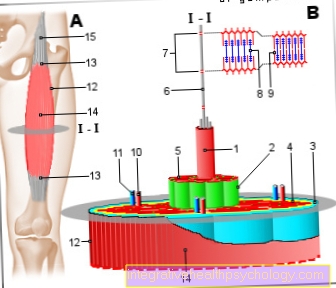
Forearm muscles
The muscles of the forearm consist of numerous muscles, some of which are smaller. To keep track of things, the muscles are divided into a
- in front (ventral) and one on the
- Rear surface (dorsal) lying muscle group.
These are each still in one
- superficial and one
- deep layer structured.
The muscles of the spoke are also separated from this.
The group of superficial anterior muscles is made up of the
- Pronator teres muscle,
- Flexor carpi radialis muscle,
- Palmaris longus muscle,
- Flexor digitorum superficialis muscle and the
- Flexor carpi ulnaris muscle together.
All of these muscles are innervated by the median nerve and originate primarily from the medial epicondyle of the upper arm.
The pronator teres muscle consists of two muscle heads and thus also arises from the coronoid process of the ulna. It starts at the middle third of the spoke and is mainly used for turning the palm of the hand in the elbow joint downwards (Pronation) responsible.
Read more on this: Forearm muscles
The flexor carpi radialis muscle extends from the medial epicondyle over the carpal bones and attaches to the second metacarpal bones. In the elbow joint it leads to
- Diffraction and
- Pronation, at the wrist he bends the hand and is for the
- Abduction, i.e. the movement towards the thumb, is responsible.
The musculus palmaris longus radiates into the muscle aponeurosis of the palm and bends the forearm in the elbow joint and the wrist.
The flexor digitorum superficialis muscle has two muscle heads. One arises from the medial epicondyle, the other from the radial head. It attaches four tendons to the metacarpal bones of the 2nd to 5th fingers.
In the elbow joint and wrist the muscle leads to flexion, and in the wrist to the movement of the hand to the little finger (Ulnar abduction). The muscle also causes flexion in the mid-metacarpal joints and a few small intermediate finger joints.
The last muscle in this group is the flexor carpi ulnaris muscle. This also consists of two heads, one originating from the medial epicondyle of the upper arm, the other from the olecranon of the ulna. It is the longest muscle and pulls to various carpal bones. The main function of the muscle is to flex the hand in the wrist. In contrast to all other muscles in this group, the flexor carpi ulnaris muscle is innervated by the ulnar nerve.
The deep layer of this front muscle group consists of the
- Pronator quadratus muscle, the
- Flexor digitorum profundus muscle and the
- Flexor pollicis longus muscle.
The pronator quadratus muscle has its origin at the distal end of the ulna on the inside of the hand and extends to the distal end of the spoke, also on the inside of the hand. This muscle lies almost squarely on the front of the forearm and causes the palm of the hand to turn downwards (Pronation). It is supplied from a branch of the median nerve.
The flexor digitorum profundus muscle arises on the front surface on about half of the ulna and attaches to the finger joints of the second to fifth fingers. This causes him to bend his fingers and wrist.
The innervation is provided by the median and ulnar nerves.
The flexor pollicis longus muscle originates from the spoke and then runs to the metacarpophalangeal joint of the thumb. Thereby it exercises its function as a flexor of the thumb. In addition, these muscles are responsible for the opposing movement of the thumb, i.e. the movement of the thumb on the palm of the hand. It is also supplied by the median nerve.
The superficial layer of the forearm muscles lying on the back is innervated by a branch of the radial nerve.
It consists of the
- Extensor digitorum muscle,
- Extensor digiti minimi muscle and dem
- Extensor carpi ulnaris muscle.
All of these muscles originate on the lateral epicondyle of the upper arm.
The extensor digitorum muscle branches out in its course and attaches to the finger joints of the 2nd to 5th finger and the wrist. This makes it the strongest flexor of the wrists and fingers.
The extensor digiti minimi muscle attaches to the little (5th) finger and stretches it. The muscle can also be absent, although this does not result in any functional restriction, since the extensor digitorum muscle then takes over the extensor function.
The extensor carpi ulnaris muscle attaches to the base of the 5th metacarpal bone and serves as a capsule amplifier. It is also used to move the wrist to the side (Ulnar abduction) responsible.
The deep layer of the back forearm muscles consists of the
- Supinator muscle,
- the abductor pollicis longus muscle,
- Extensor pollicis brevis and longus muscles, as well as the
- Extensor indicis muscle.
All muscles are innervated by the radial nerve or by branches of this nerve.
The supinator muscle, as its name suggests, is involved in the supination of the hand. That means he turns the palm of the hand upwards. It has its origin in the lateral epicondyle of the upper arm and runs from there obliquely to the forearm axis on the front surface of the spoke (radius).
The abductor pollicis longus muscle originates from the back of the ulna and radius and attaches to the base of the thumb. In the wrist it causes a flexion and splaying of the radius towards the radius (after radial). However, its main function is to stretch (Extension) and splay (Abduction) of the thumb saddle joint.
The abductor pollicis brevis muscle arises on the back surface of the spoke and attaches to the base of the thumb. It thus has the same function as the abovementioned abductor pollicis longus muscle.
The extensor pollicis longus muscle originates from the back of the ulna and extends from there to the base of the thumb. Here it leads to stretching (Extension) and pre-accession (Adduction) of the thumb.
The last muscle in this group is the extensor indicies muscle, which extends the index finger. It arises from the back of the spoke and attaches to the base of the index finger.
Joints of the forearm
They are in the elbow joint
- distant (distal) End of the humerus, as well as the
- body-hugging (proximal) Ends of cubit (Ulna) and spoke (radius) in an articulated connection.
The three compartments each form a joint, whereby the elbow joint consists of three partial joints.
The humerus stands with both the
- Cubit (Articulatio humeroulnaris) and with the
- Spoke (Articulatio humeroradialis) in an articulated connection.
The third joint is formed between the two proximal ends of the ulna and radius that are close to the body (articulatio radioulnaris proximalis).
Through these different joints there is one
- Diffraction (Flexion) and
- Elongation (Extension) as well as the
- Rotation of the forearm or palm upwards (Supination) and down (Pronation) possible.
The distant (distal) Ellen's spoke joint (Articulatio radioulnaris distalis) also allows the radius to rotate around the ulna and thus the palm of the hand to move upwards (Supination) and down (Pronation).
In addition, the ulna and radius together with the (proximal) Carpal bones the proximal wrist.
This enables
- Diffraction (Flexion) and
- Elongation (Extension), as well as the
- Splay to ulna (Ulnar abduction) and to the spoke (Radial abduction).
Vessels of the forearm
Arteries
The forearm receives its supply primarily from the large arm artery coming from the upper arm (Brachial artery). In the crook of the elbow this splits into the
- Spoke side (Radial artery) and the
- Ellside (Ulnar artery) on.
These in turn divide into numerous smaller and larger branches, which then supply the forearm and muscles and ultimately flow into the vascular network of the hand.
Veins
From the hand goes on the
- The cephalic vein and on the spoke side
- The basilic vein emerges as superficial veins on the ulnar side.
The deep large veins run with the arteries and are named like them.
The intermediate vena antebrachii also runs as large superficial veins. It runs relatively centrally on the front of the forearm. In the crook of the elbow there is a connection between the
- external cephalic vein and the
- inner basilic vein.
This connection is known as the median cubital vein. Since it is easily visible and palpable from the outside, it is often used for intravenous injections or for taking blood.
Nerves of the forearm
The forearm muscles are mainly made up of nerves from the nerve branch plexus of the arm (Brachial plexus) innervated.
These nerves take their course over the upper arm and then innervate the forearm
- Skin and
- Musculature.
The radial nerve primarily innervates the muscles of the back of the forearm and the extensor muscles of the forearm.
Furthermore, sensitive skin nerves branch off from it, which parts of the
- Thumb and des
- Sensitively care for the back of the hand.
The median nerve innervates almost all of the flexor muscles of the forearm, and some sensitive fibers also come from this nerve. These supply the skin on the inside of the hand between the thumb and ring finger.
The ulnar nerve, on the other hand, innervates very few muscles:
- the flexor carpi ulnaris muscle and
- the flexor digitorum profundus muscle.
It also innervates the skin on the inside of the hand and the back of the hand between the ring finger and the edge of the hand.
Recommendations from our editorial team
- Vascular supply arm
- Forearm pain
- Wrist pain
- Wrist pain
- Broken wrist
- Wrist osteoarthritis
- Broken arm child